Lacertus Syndrome
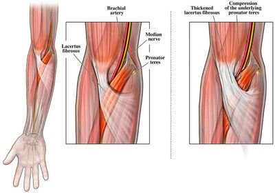
Lacertus syndrome is a chronic exertional compartment syndrome (CECS) of the forearm, distinct from exertional compartment syndrome in the lower extremity. It involves the compression of the pronator teres muscle by the lacertus fibrosis, often seen in athletes engaged in overhead throwing activities. This syndrome exhibits unique anatomical, clinical, and diagnostic features compared to traditional CECS.
Anatomy and Pathophysiology

Lacertus syndrome was first described by George Bennett in 1959 as a condition affecting overhead throwing athletes. It is caused by compression of the pronator teres muscle by the lacertus fibrosis, leading to disabling pain in the proximal volar forearm. This compression can also involve the median nerve, adding to the symptom complexity. The pain typically starts as an ache at the medial elbow during exertion, resolving after several hours of rest. Without cessation of activity, the symptoms can intensify and persist longer. The delayed onset and resolution with rest are characteristic of exertional compartment syndrome but differ from other proximal forearm neuropathies.
The median nerve runs along the medial side of the brachial artery, situated between the biceps brachii and the brachialis muscle. It courses under the ligament of Struthers and often gives a branch to the pronator teres just distal to the elbow. The nerve remains medial to the biceps tendon and brachial artery throughout its course and is located deep to both the lacertus fibrosis (bicipital aponeurosis) and the pronator teres muscle. The lacertus fibrosis is not the primary fascia of the superficial volar compartment but acts as a constricting band around the pronator teres. This constriction can lead to elevated pressure within the pronator teres during muscle exertion, without elevating the pressure in the entire forearm compartment.
Clinical Features

Patients with lacertus syndrome typically present with increasing pain and a sense of fullness in the flexor-pronator muscle group with exertion. The symptoms, including intermittent median nerve compression, resolve after rest, which can last from several hours to days. The most commonly affected individuals are baseball pitchers and American football quarterbacks, due to the repetitive nature of their arm movements, which predisposes them to pronator teres compression.
Lacertus syndrome shares symptoms with other elbow pathologies, such as cubital tunnel syndrome, ulnar neuritis, pronator syndrome, anterior interosseous nerve syndrome, and medial epicondylitis. Hence diagnosis can be difficult.
If lacertus syndrome is suspected, the patient should be examined after exertion to detect swelling or contour changes in the flexor-pronator mass, as well as reproduction of pain with wrist flexion, pronation, and elbow flexion.
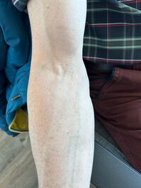
The lacertus notch is present in the majority of cases.
Investigations
Unlike static neuropathies like pronator syndrome, the dynamic nature of lacertus syndrome makes it harder to diagnose using conventional methods. Invasive pressure measurements, similar to those used for CECS in the legs, have been employed in the past, but they carry risks of injury to surrounding structures. Recent advances, including pre- and post-exercise MRI, have provided a less invasive and reliable diagnostic method.[1]
Treatment
Surgical release of the lacertus fibrosis has been shown to provide excellent outcomes for lacertus syndrome.
Literature Review
- Reviews from the last 7 years: review articles, free review articles, systematic reviews, meta-analyses, NCBI Bookshelf
- Articles from all years: PubMed search, Google Scholar search.
- TRIP Database: clinical publications about evidence-based medicine.
- Other Wikis: Radiopaedia, Wikipedia Search, Wikipedia I Feel Lucky, Orthobullets,


