Cervical Pain Maps
Facetogenic Pain
Symptomatic Patients
Cooper et al (C1/2 to C6/7)
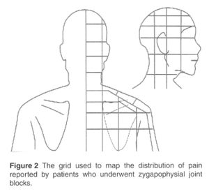
- Cervical Pain Maps for Medial Branch Blocks
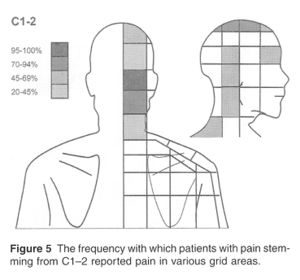
C1-2
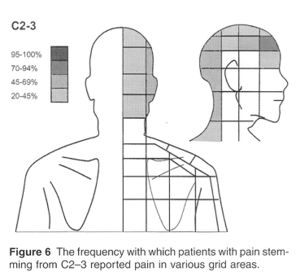
C2-3
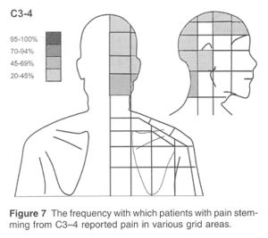
C3-4
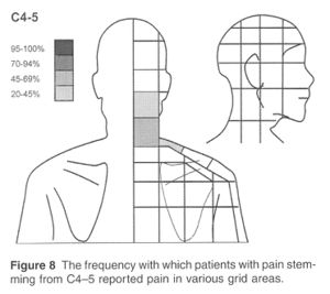
C4-5
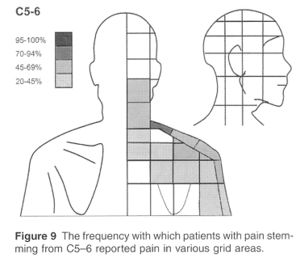
C5-6
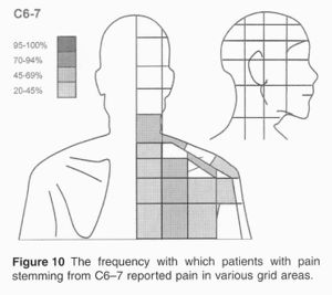
C6-7
C0-3
Blank Grid
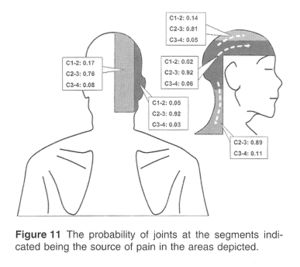
Probabilities C1-C4
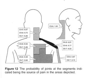
Probabilities C4-C7


Images Copyright © 2007 American Academy of Pain Medicine
Full Image: File:Cervical Pain Maps Grid.jpg [1]
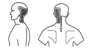
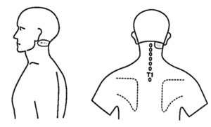
Fukui et al (C0/1 to C7/T1)
61 symptomatic patients with suspected facet joint pain. Pain reproduced by injection of contrast medium into the joints or by electrical stimulation of the dorsal rami. Below shows the main pain distributions from C0-1 to C7/T1, and the dorsal rami C3 to C7[2]

| Joint | Pain Referral |
|---|---|
| C0/1 | Occipital region (30%), upper posterolateral cervical region (100%) |
| C1/2 | Occipital region (20%) and upper posterolateral cervical region (100%). |
| C2/3 | Upper posterior cervical region (64%), occipital region (50%), and upper posterolateral cervical region (50%) |
| C3/4 | Upper posterior cervical region (76%), middle posterior cervical region (52%), and occipital region (38%) |
| C4/5 | Lower posterior cervical region (76%), middle posterior cervical region (54%), and suprascapular region (43%) |
| C5/6 | suprascapular region (50%),lower posterior cervical region (46%), superior angle of the scapula (35%), middle posterior cervical region (15%), and shoulder joint (11%) |
| C6/7 | superior angle of the scapula (48%), mid-scapular region (41%), lower posterior cervical region (33%), shoulder joint (15%),and suprascapular region (11%). |
| C7/T1 | Mid-scapular region (86%) and superior angle of the scapula (28%) |
Normal Volunteers
Dwyer et al (C2/3 to C6/7)
Pain maps with noxious stimulation of cervical facet joints in volunteers,[3] which was then validated against symptomatic patients.[4]

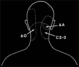
Dreyfuss et al (C0/1 and C1/2)
The pain referral patterns of C0-1 and C1-2 were evaluated in 5 normal volunteers.[5]
C0-1
C1-2


Discogenic Pain
Disc pain has very similar referral patterns to zygapophyseal joint pain. Below is the pattern of pain provoked by discography at each cervical level: C2–C3 (A), C3–C4 (B), C4 –C5 (C), C5–C6 (D), and C6 –C7 (E).

Radicular Pain
- Main article: Cervical Radicular Pain
![Based off data from Rainville et al. The asterix indicates the only area where there is a statistical difference, with impaired in the distal radial aspect of the dorsal forearm more common in C6 than C7 radiculopathy[6]](/w/img_auth.php/thumb/1/1d/C6_and_C7_radicular_pain.png/500px-C6_and_C7_radicular_pain.png)
Based off data from Rainville et al. The asterix indicates the only area where there is a statistical difference, with impaired in the distal radial aspect of the dorsal forearm more common in C6 than C7 radiculopathy[6]
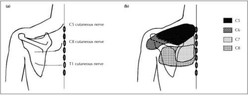
(a)C5 and C8 cutaneous nerves traverse up and down the spine of the scapula, respectively, whereas the courses of C6 and C7 cutaneous nerves are not found.
(b) The site of radicular pain involving the C6 root overlaps with that of the C5 root, and also involves the posterior deltoid.
![Based off data from Rainville et al. The asterix indicates the only area where there is a statistical difference, with impaired in the distal radial aspect of the dorsal forearm more common in C6 than C7 radiculopathy[6]](/wiki/File:C6_and_C7_radicular_pain.png)

Interspinous Ligaments
Referred pain patterns from noxious stimulation of the cervical interspinous ligaments. He described injecting into the midline and just off centre into the interspinous ligaments.[7]

Trigger Points
This is a clinical entity, and is very difficult to reproduce in a research setting.
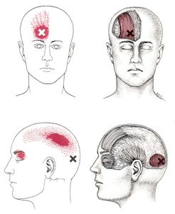
Occipitalis, Frontalis
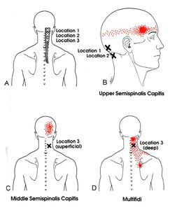
Semispinalis Capitis, Multifidi
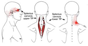
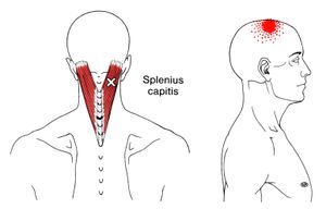
Splenius Capitis, Splenius Cervicis
Splenius Capitis, Splenius Cervicis 2
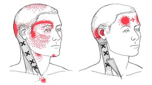
Sternocleidomastoid
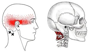
Suboccipital Group
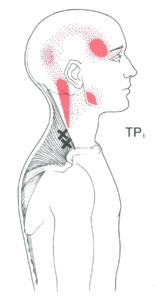
Trapezius 1
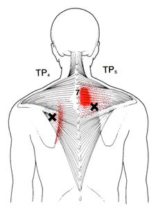
Trapezius 2
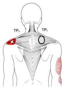
Trapezius 3


See Also
References
- ↑ Cooper et al.. Cervical zygapophysial joint pain maps. Pain medicine (Malden, Mass.) 2007. 8:344-53. PMID: 17610457. DOI.
- ↑ Fukui et al.. Referred pain distribution of the cervical zygapophyseal joints and cervical dorsal rami. Pain 1996. 68:79-83. PMID: 9252002. DOI.
- ↑ Dwyer et al.. Cervical zygapophyseal joint pain patterns. I: A study in normal volunteers. Spine 1990. 15:453-7. PMID: 2402682. DOI.
- ↑ Aprill et al.. Cervical zygapophyseal joint pain patterns. II: A clinical evaluation. Spine 1990. 15:458-61. PMID: 2402683. DOI.
- ↑ Dreyfuss et al.. Atlanto-occipital and lateral atlanto-axial joint pain patterns. Spine 1994. 19:1125-31. PMID: 8059267. DOI.
- ↑ Rainville et al.. Exploration of sensory impairments associated with C6 and C7 radiculopathies. The spine journal : official journal of the North American Spine Society 2016. 16:49-54. PMID: 26253986. DOI.
- ↑ Kellgren. On the distribution of pain arising from deep somatic structures with charts of segmental pain areas. Clinical Science. 1939.

