Category:License templates
From WikiMSK
Pages in category 'License templates'
The following 10 pages are in this category, out of 10 total.
Media in category 'License templates'
The following 200 files are in this category, out of 647 total.
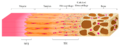
(previous page) (next page)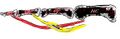 Finger flexor tendons.jpg 578 × 186; 48 KB
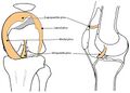
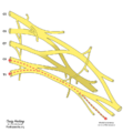
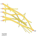
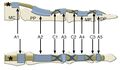 Finger pulley system.jpg 763 × 445; 47 KB
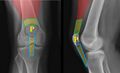
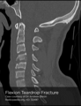 Flexion teardrop.png 450 × 586; 124 KB
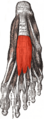 Flexor digitorum brevis.png 297 × 800; 79 KB
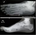
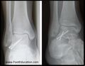
 Foot and Ankle Radiograph Normal.jpg 647 × 761; 59 KB
 Fracture base of 5th metacarpal.jpg 768 × 630; 62 KB
 Fracture shaft 5th metacarpal.jpg 368 × 370; 34 KB
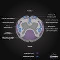
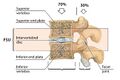 Functional spinal unit.jpg 1,295 × 863; 144 KB
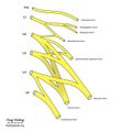
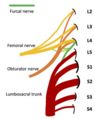 Furcal nerve.png 277 × 348; 54 KB
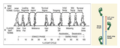 Gait diagram.png 1,380 × 548; 998 KB
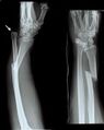 Galeazzi fracture.jpg 239 × 300; 10 KB
 Ganglion cyst.jpg 384 × 470; 45 KB
 Ganglion impar fluoroscopy.PNG 338 × 332; 100 KB
 Ganglion volar wrist MRI.png 298 × 300; 81 KB
 Gartland type 3 fracture.jpg 1,085 × 469; 59 KB
 Genomic sequencing history.jpg 1,107 × 741; 141 KB
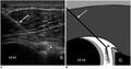 GHJ injection under US.jpg 792 × 418; 41 KB
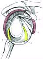 Glenohumeral joint capsular structures.jpg 717 × 974; 94 KB
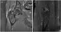 Gluteus medius MRI high grade partial thickness tear.jpg 688 × 368; 85 KB
 Gluteus medius tear resisted internal rotation test.jpg 624 × 200; 36 KB
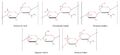 Glycosaminoglycans.jpg 3,753 × 1,716; 93 KB
 Golgi tendon organ.png 700 × 240; 30 KB
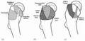
 GON and TON emergences.png 1,500 × 911; 519 KB
 GON and TON ultrasound.png 950 × 494; 480 KB
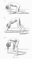 Gowers sign.jpg 393 × 720; 45 KB
 Grant MCP.png 768 × 1,074; 258 KB
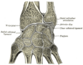
 Gray186 hyoid bone.jpg 468 × 321; 60 KB
 Gray331 Elbow joint capsule anterior.png 252 × 550; 32 KB
 Gray333 Annular ligament radius from above.png 404 × 388; 35 KB
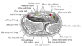
 Gray336 coronal section wrist joints.png 550 × 430; 48 KB
 Gray421.png 550 × 312; 26 KB
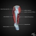
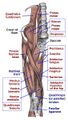 Gray430 anterior lower limb muscles.jpg 678 × 1,192; 182 KB
 Gray545.png 500 × 623; 98 KB
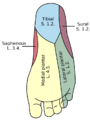 Gray834 plantar foot sensation.png 578 × 768; 69 KB
 Greater trochanter anatomy facets insertions and bursae.jpg 768 × 369; 48 KB
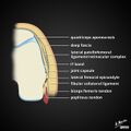
 Greater trochanter facets.png 425 × 566; 126 KB
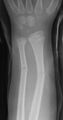 Greenstick fractures ulna and radius.jpg 158 × 300; 6 KB
 Hamate fracture on CT.jpg 300 × 300; 10 KB
 Hamate.jpg 181 × 300; 13 KB
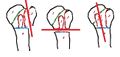
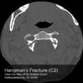 Hangman-fracture.png 450 × 450; 88 KB
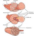 Hierarchical-structure-of-skeletal-muscle.jpg 1,435 × 1,482; 220 KB
 High importance.png 50 × 50; 1 KB
 Hindfoot alignment radiograph.jpg 634 × 1,100; 113 KB
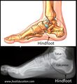 Hindfoot anatomy.jpg 824 × 900; 88 KB
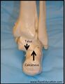 Hindfoot posterior.jpg 697 × 900; 58 KB
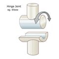 Hinge joint.jpg 704 × 704; 105 KB
 Hip abductor strength testing.jpg 609 × 484; 96 KB
 Hip anatomy cadaver.jpg 960 × 720; 96 KB
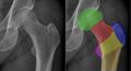
 Hip AP view radiographic signs.png 1,039 × 260; 129 KB
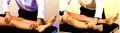
 Hip flexion test.jpg 1,024 × 768; 102 KB
 Hip fracture screws xr.jpg 425 × 216; 13 KB
 Hip injection anterior longitudinal approach.jpg 400 × 626; 35 KB
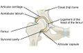
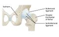
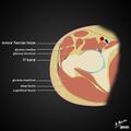
 Hip joint anterior view.jpg 1,092 × 700; 85 KB
 Hip joint frontal section.jpg 1,090 × 700; 118 KB
 Hip joint injection anterior longitudinal approach ultrasound.jpg 640 × 491; 48 KB
 Hip joint injection anterolateral approach ultrasound.jpg 640 × 626; 56 KB
 Hip Joint Injection Ultrasound.png 1,029 × 1,121; 251 KB
 Hip joint posterior view.jpg 1,096 × 700; 86 KB
 Hip log roll test.jpg 1,326 × 367; 69 KB
 Hip neck shaft angle of inclination.jpg 686 × 197; 51 KB
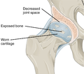 Hip osteoarthritis illustration.png 898 × 791; 319 KB
 Hip pain pointing.jpg 539 × 213; 26 KB
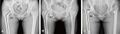
 Hip xr anatomy.jpg 1,957 × 1,053; 208 KB
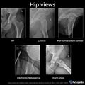 Hip-views-1.jpeg 1,600 × 1,600; 776 KB
 HIT trial results.jpg 1,500 × 688; 63 KB
 Homunculus.jpg 1,599 × 1,593; 84 KB
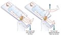 Hoover Sign.jpg 595 × 340; 51 KB
 Human skeleton front.png 310 × 599; 83 KB
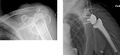
 Humeral head four part fracture.jpg 277 × 323; 11 KB
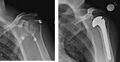 Humeral head fracture and hemiplasty.jpg 699 × 361; 33 KB
 Humeral head fracture and reverse hemiarthroplasty.jpg 610 × 278; 27 KB
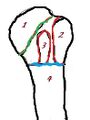 Humeral head osseous segments.jpg 197 × 262; 9 KB
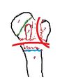 Humeral head three part fracture.jpg 276 × 373; 14 KB
 Humeral head two part fractures.jpg 649 × 306; 32 KB
 Humeral head xray.jpg 879 × 809; 48 KB
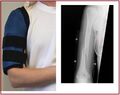 Humerus fracture bracing.jpg 1,519 × 1,197; 136 KB
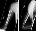 Humerus fracture fixation.jpg 1,047 × 900; 83 KB
 Humerus fracture radial nerve.jpg 1,861 × 771; 106 KB
 Humerus oblique fracture.jpg 672 × 1,101; 60 KB
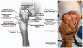
 Humerus shaft and distal anatomy.jpg 1,747 × 869; 113 KB
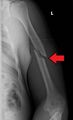
 Humerus shaft fracture.jpg 178 × 450; 16 KB
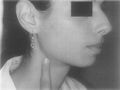 Hyoid syndrome tenderness.jpg 657 × 493; 66 KB
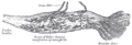
 Hypaxial and epaxial muscles in a fish.png 220 × 211; 17 KB
 Hyperpathia increased DT decreased PT allodynia.png 3,420 × 1,976; 70 KB
 Hyperpathia increased DT decreased PT hyperalgesia.png 3,420 × 1,976; 69 KB
 Hyperpathia increased DT increased PT hyperalgesia.png 3,420 × 1,976; 72 KB
 Hyperpathia increased DT normal PT hyperalgesia.png 3,420 × 1,976; 68 KB
 Hyperpathia unmasking.png 3,420 × 1,976; 85 KB
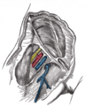
 Iliac fossa dissection.png 451 × 524; 139 KB
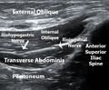 Ilioinguinal nerve block fascial plane.jpg 624 × 516; 182 KB
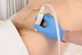 Ilioinguinal nerve injection probe position.jpg 412 × 274; 66 KB
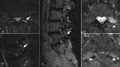
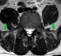 Iliolumbar ligament on MRI.PNG 536 × 496; 223 KB
 Iliotibial-band-hip-illustration.jpg 640 × 640; 38 KB
 Iliotibial-band-illustration.jpg 640 × 640; 31 KB
 Iliotibial-band-knee-illustration.jpg 640 × 640; 38 KB
 Iliotibial-band-origins-illustration.jpg 640 × 640; 31 KB
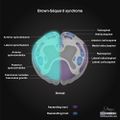 Incomplete-spinal-cord-syndromes-illustrations brown sequard.jpg 500 × 500; 53 KB
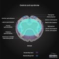 Incomplete-spinal-cord-syndromes-illustrations central.jpg 500 × 500; 52 KB
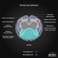 Incomplete-spinal-cord-syndromes-illustrations dorsal.jpg 500 × 500; 53 KB
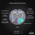 Incomplete-spinal-cord-syndromes-illustrations posterior.jpg 500 × 500; 56 KB
 Incomplete-spinal-cord-syndromes-illustrations ventral.jpg 500 × 500; 56 KB
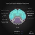
 Incomplete-spinal-cord-syndromes-illustrations.jpg 500 × 500; 54 KB
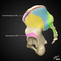
 Innominate bony landmarks.jpg 974 × 700; 317 KB
 Instability factor.jpg 2,241 × 1,145; 96 KB
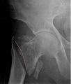 Intertrochanteric hip fracture.jpg 368 × 433; 18 KB
 Intraabdominal pressure test.jpg 436 × 281; 20 KB
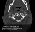 Jefferson.png 450 × 418; 69 KB
 Jones fracture 5th metatarsal.jpg 909 × 898; 64 KB
 Keinbocks MRI.png 234 × 300; 67 KB
 Keinbocks xray.png 232 × 300; 65 KB
 Knee blood supply.png 500 × 453; 40 KB
 Knee Bursae.jpg 1,121 × 899; 116 KB
 Knee joint.png 512 × 512; 60 KB
- Knee plica inspection.mp4 ; 4.42 MB
 Knee plicae.jpg 800 × 570; 140 KB
 Knee sunrise view.jpg 685 × 329; 20 KB
 Knee xray.jpg 1,430 × 869; 174 KB
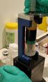 L-PRP extraction.jpg 372 × 642; 84 KB
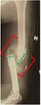
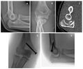
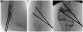 Lateral condyle fracture surgical reduction.jpg 844 × 328; 33 KB
 Lateral hip illustration and cadaver.jpg 624 × 360; 48 KB
 Lateral process fracture surgery.jpg 885 × 689; 62 KB
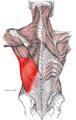 Latissimus dorsi.jpg 640 × 1,009; 168 KB
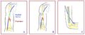
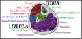 Leg compartments.jpg 946 × 461; 93 KB
 Lock-green.png 100 × 159; 5 KB
 Loeys-Dietz syndrome manifestations.jpg 1,660 × 2,079; 168 KB
 Loss of rotational anatomic alignment proximal phalanx fracture.jpg 300 × 251; 14 KB
 LTN and SAN palsies.jpg 852 × 433; 56 KB
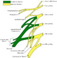
 Lumbar and Sacral Plexus.jpg 1,802 × 1,194; 325 KB
 Lumbar facet joints MRI oedema.jpg 1,032 × 577; 98 KB
 Lumbar medial branch blocks fluoroscopy left L3-5.jpg 743 × 725; 81 KB
 Lumbar Plexus Hacking.jpeg 630 × 630; 52 KB
 Lumbar Plexus.png 483 × 488; 65 KB
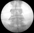
 Lumbar Spine AP Radiograph Normal.png 263 × 524; 112 KB
 Lumbar Spine L3 Lateral Normal.png 1,104 × 842; 641 KB
 Lumbar Spine L4 and L5 AP Radiograph Normal.png 852 × 820; 546 KB
 Lumbar Spine L4-5 Lateral Normal.png 1,020 × 860; 702 KB
 Lumbar Spine Lateral Radiograph Normal.png 322 × 508; 104 KB
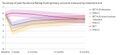
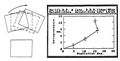
 Lumbar TFI success rates Ghahreman 2010 chart.png 1,101 × 547; 57 KB
 Lumbar-interbody-fusion.png 386 × 380; 139 KB
 Lumbar-neuroforaminal-stenosis-mild-disc.jpg 686 × 686; 53 KB
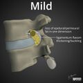 Lumbar-neuroforaminal-stenosis-mild-ligamentum-flavum.jpg 686 × 686; 53 KB
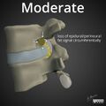 Lumbar-neuroforaminal-stenosis-moderate.jpg 686 × 686; 53 KB
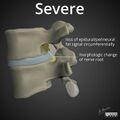 Lumbar-neuroforaminal-stenosis-severe.jpg 686 × 686; 54 KB
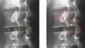 Lumbar-spine-annotated-oblique-projection.jpg 1,031 × 588; 315 KB
 Lunate.jpg 181 × 300; 13 KB
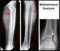 Maisonneuve fracture.jpg 973 × 829; 82 KB
 Mallet finger patient.jpg 360 × 235; 62 KB
 Mallet finger.jpg 1,054 × 333; 28 KB
 Mason type 1 radial head fracture.jpg 557 × 400; 42 KB
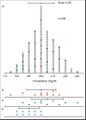 Mean and SD example.jpg 739 × 1,027; 71 KB
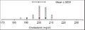 Mean and SEM example.jpg 737 × 263; 21 KB
 Medial antebrachial cutaneous nerve.png 768 × 768; 154 KB
 Medial epicondyle fracture.jpg 827 × 689; 67 KB
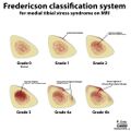 Medial tibial stress Fredericson grading.jpg 1,024 × 1,024; 115 KB
 Median nerve.png 768 × 768; 168 KB
 Metacarpal bones.jpg 181 × 300; 13 KB
 Metacarpal fracture surgery.jpg 661 × 740; 41 KB
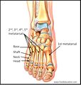 Metatarsal anatomy.jpg 805 × 847; 82 KB
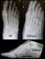 Metatarsals on xray.jpg 676 × 895; 64 KB
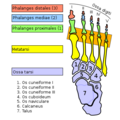 Metatarsophalangeal joints with latin names.png 375 × 376; 45 KB
 Midshaft clavicle fracture.jpg 954 × 541; 52 KB
 Monoarticular arthralgia algorithm.png 837 × 661; 27 KB
 Monteggia fracture.jpg 240 × 559; 22 KB
 Morphine chemical structure in 3D.png 742 × 553; 11 KB
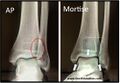 Mortise view ankle.jpg 1,090 × 750; 118 KB
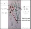 Mortons neuroma plantar nerves.jpg 957 × 892; 93 KB
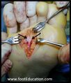 Mortons neuroma surgery.jpg 536 × 628; 53 KB
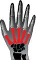
 Motor actions of hand musles innervated by median nerve.jpg 800 × 262; 169 KB
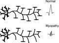 Motor units in myopathies.jpg 751 × 548; 44 KB
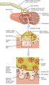 Motor-end-plate.jpg 1,462 × 2,548; 279 KB
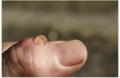 Mucous cyst of the thumb.png 300 × 196; 29 KB
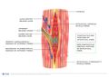 Muscle Spindle.jpg 2,048 × 1,448; 188 KB
 Muscle-fibre.jpg 801 × 642; 105 KB
 Muscles plantar foot first layer Sobo.jpg 719 × 1,010; 110 KB
 Muscles plantar foot Gray444.png 330 × 800; 59 KB
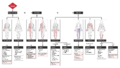
 Muscular dystrophy weakness patterns.jpg 607 × 443; 66 KB
 Musculusconstrictorpharyngismedius.jpg 360 × 599; 46 KB
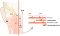
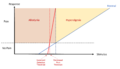
 Myotendinous junction and enthesis combined.png 850 × 323; 84 KB
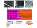 Myotendinous junction structure.png 850 × 648; 137 KB
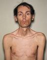 Myotonic dystrophy patient.JPG 162 × 208; 5 KB
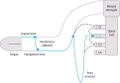 Neck tongue syndrome fibres.jpg 484 × 333; 27 KB
 Needle bend.jpg 1,340 × 282; 35 KB
 Needle bevel direction.jpg 2,449 × 1,402; 169 KB
 Negative ulnar variance.png 396 × 272; 101 KB
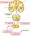 Nerve impulses transduction to perception.png 668 × 790; 129 KB
 Neuroanatomy-of-the-lumbar-discovertebral-complex.png 702 × 607; 195 KB
 Neurogenic inflammation.png 850 × 513; 95 KB
 Neuropathic heel ulcer diabetic.jpg 750 × 563; 40 KB
 Neurotransmitters CWP.jpg 1,280 × 720; 150 KB
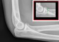
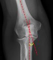
 Normal elbow xray.jpg 674 × 293; 18 KB
 Normal-elbow-carrying-angle.jpg 474 × 537; 123 KB
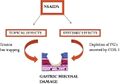 NSAID induced GIT effects.jpg 591 × 415; 27 KB
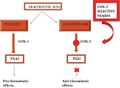 NSAID induced thrombosis.jpg 591 × 433; 24 KB
 Numbness algorithm.png 2,955 × 1,672; 179 KB
 Nzamm-logo-long.png 1,504 × 413; 59 KB
 NZCMM logo.png 512 × 512; 88 KB


