Cervical Intervertebral Discs: Difference between revisions
No edit summary |
No edit summary |
||
| Line 8: | Line 8: | ||
The cervical disc is not a small lumbar disc, but is structurally unique. | The cervical disc is not a small lumbar disc, but is structurally unique. | ||
== Overview == | |||
{{Cervical spine anatomy and biomechanics}} | |||
== Structure == | == Structure == | ||
The annulus is not concentric, but rather is only well developed at the anterior aspect. The collagen fibres form a thick crescentic mass anteriorly and taper laterally towards the uncinate processes. There also isn't the criss-cross pattern of collagen fibres like in the lumbar discs. Rather, the fibres converge upwards towards the anterior end of the upper vertebra. When viewed front-on, the annulus is shaped like an inverted "V", with the fibres converging towards the midline, and the apex of the V pointing to the axis of rotation. In essence, the cervical annulus is more like an anterior interosseous ligament and is not load bearing.<ref name=":0">{{#pmid:10209789}}</ref> | The annulus is not concentric, but rather is only well developed at the anterior aspect. The collagen fibres form a thick crescentic mass anteriorly and taper laterally towards the uncinate processes. There also isn't the criss-cross pattern of collagen fibres like in the lumbar discs. Rather, the fibres converge upwards towards the anterior end of the upper vertebra. When viewed front-on, the annulus is shaped like an inverted "V", with the fibres converging towards the midline, and the apex of the V pointing to the axis of rotation. In essence, the cervical annulus is more like an anterior interosseous ligament and is not load bearing.<ref name=":0">{{#pmid:10209789}}</ref> | ||
Revision as of 08:49, 12 June 2022

| |
| Cervical Intervertebral Discs | |
|---|---|
| Primary Type | Cartilaginous Joint |
| Secondary Type | Symphysis |
| Bones | |
| Ligaments | |
| Muscles | |
| Innervation | |
| Vasculature | |
| ROM | |
| Volume | |
| Conditions | |
The cervical disc is not a small lumbar disc, but is structurally unique.
Overview
| C0-1 | C1-2 | C2-7 | |
|---|---|---|---|
| Anatomy | 1. Superior facets of C0 (Atlas): 28° in sagittal and transverse planes
2. No disc |
1. C1 has convex facet joint surface (allow C1 facet to slide in AP direction over C2)
2. No disc 3. Transverse lig : prevent dense posterior dislocation 4. Alar lig: limiting rotation |
Anatomy structure grossly similar.
1. 2x uncovertebral joints (joints of Luschka) at each segments. They are not true synovial joints (clefts at the lateral edges of IVD between uncinated process inferiorly, and the edge of the vertebral body above) 2. 1 disc between each vertebra body. 3. 2 facet joints: at 45° inclination to horizontal plan (pure rotation, side bending is limited) |
| Biomechanics |
|
Rotation: 45° both directions.
(Rotation in full flexion is C1-2 rotation) |
|
Structure
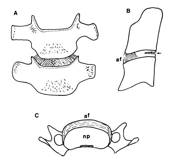
The annulus is not concentric, but rather is only well developed at the anterior aspect. The collagen fibres form a thick crescentic mass anteriorly and taper laterally towards the uncinate processes. There also isn't the criss-cross pattern of collagen fibres like in the lumbar discs. Rather, the fibres converge upwards towards the anterior end of the upper vertebra. When viewed front-on, the annulus is shaped like an inverted "V", with the fibres converging towards the midline, and the apex of the V pointing to the axis of rotation. In essence, the cervical annulus is more like an anterior interosseous ligament and is not load bearing.[2]
Posteriorly there is only a thin layer of paramedian, vertically oriented collagen fibres. The posterior aspect of the disc is really only covered by the posterior longitudinal ligament.[2] The reason there is no posterior annulus is because it would impede rotation along the oblique coronal plane.
Age Changes
At birth the nucleus pulposus is relatively small, but persists until the second decade. Following that it gradually disappears as it becomes fibrocartilage. This results in cervical discs being harder and drier than lumbar discs. [3]
In degenerative change cervical discs form internal cracks and fissures, and slowly develop fibrocartilaginous bulges and osteophytes. It is normal to have transverse fissures on the posterior aspects of the discs. These initially appear in childhood in the uncovertebral regions, and with age extend medially, finally forming transverse clefts by the third decade of life.[3] These transverse fissures facilitate axial rotation,[1] they are effectively joint spaces between the vertebral bodies.
Innervation
The nucleus pulposus is not innervated. The outer annulus fibrosus is innervated anteriorly by the vertebral nerve, and posterolaterally by the sinuvertebral nerve.
References
- ↑ 1.0 1.1 Bogduk & Mercer. Biomechanics of the cervical spine. I: Normal kinematics. Clinical biomechanics (Bristol, Avon) 2000. 15:633-48. PMID: 10946096. DOI.
- ↑ 2.0 2.1 Mercer & Bogduk. The ligaments and annulus fibrosus of human adult cervical intervertebral discs. Spine 1999. 24:619-26; discussion 627-8. PMID: 10209789. DOI.
- ↑ 3.0 3.1 Bogduk N. Degenerative joint disease of the spine. Radiol Clin North Am. 2012 Jul;50(4):613-28. doi: 10.1016/j.rcl.2012.04.012. PMID: 22643388.

