◒
De Quervain Injection: Difference between revisions
From WikiMSK
No edit summary |
|||
| Line 13: | Line 13: | ||
==Anatomy== | ==Anatomy== | ||
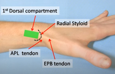
The APL and EPB usually run together in the first dorsal compartment. The tendons can often be seen with the thumb held in resisted extension. They can also be palpated at the base of the 1st metacarpal. | *The APL and EPB usually run together in the first dorsal compartment. | ||
*The tendons can often be seen with the thumb held in resisted extension. | |||
*They can also be palpated at the base of the 1st metacarpal. | |||
*Anatomic variation: septum with two sub compartments (24-76% in cadaver studies). Failure can occur if failure to inject into compartment or only one sub compartment. | |||
[[File:First Dorsal Compartment.PNG|400px]] | [[File:First Dorsal Compartment.PNG|400px]] | ||
| Line 22: | Line 25: | ||
==Technique== | ==Technique== | ||
[[File:De Quervain Ultrasound Injection.PNG|400px|thumb|Long axis injection. From left to right: needle, APB, EPL.]] | |||
*Ultrasound guided is preferred with greater clinical improvement, and allows the identification of subcompartment anatomical variation <ref>McDermott JD, Ilyas AM, Nazarian LN, et al. Ultrasound-guided injections for de Quervain's tenosynovitis. Clin Orthop Relat Res. 2012;470:1925–31.</ref><ref>Jeyapalan K, Choudhary S. Ultrasound-guided injection of triamcinolone | |||
and bupivacaine in the management of de Quervain’s disease. Skelet Radiol. 2009;38:1099–103.</ref><ref>Zingas C, Failla JM, Van Holsbeeck M. Injection accuracy and clinical | |||
relief of de Quervain’s tendinitis. J Hand Surg Am. 1998;23:89</ref> | |||
*Position: Ulnar side of hand resting on surface with thumbheld in slight flexion | *Position: Ulnar side of hand resting on surface with thumbheld in slight flexion | ||
| Line 35: | Line 42: | ||
===Ultrasound Guided=== | ===Ultrasound Guided=== | ||
* Identify: APL and APB tendons in sagittal, retinaculum, radial styloid in transverse | * Identify: APL and APB tendons in sagittal, retinaculum, radial styloid in transverse | ||
* | * Stand off gel recommended | ||
** | * Can be done long axis or short axis. Transverse view is best with the needle entering the sheath while in plan with the transducer. | ||
* | * Avoid the superficial branch of the radial nerve | ||
* Inject within the tendon sheath. | |||
==Complications== | ==Complications== | ||
Revision as of 19:30, 30 June 2020
This article is still missing information.
| De Quervain Injection | |
|---|---|
| Indication | De Quervain Tendinopathy |
| Syringe | 1mL |
| Needle | 25G 16mm |
| Steroid | 10-20mg triamcinolone |
| Local | 0.75mL 2% lidocaine |
| Volume | 1mL |
Background
Injection for De Quervain Tendinopathy.
Anatomy
- The APL and EPB usually run together in the first dorsal compartment.
- The tendons can often be seen with the thumb held in resisted extension.
- They can also be palpated at the base of the 1st metacarpal.
- Anatomic variation: septum with two sub compartments (24-76% in cadaver studies). Failure can occur if failure to inject into compartment or only one sub compartment.
Indications
Contraindications
Technique
- Ultrasound guided is preferred with greater clinical improvement, and allows the identification of subcompartment anatomical variation [1][2][3]
- Position: Ulnar side of hand resting on surface with thumbheld in slight flexion
Non-Ultrasound Guided
- Identify: Radial styloid, the APB and EPL tendons, and the gap between them.
- Injection site
- Usual site: is between 5-10mm proximal to the tip of the radial styloid, between the two tendons, through the retinaculum, within the sheath.
- Alternative site in very thin patients: inject distal to the retinaculum, 5mm distal to the radial styloid (due to limited subcutaneous tissue), then advance the needle proximally while injecting
- Insert needle perpendicularly into the gap then slide proximally between the tendons (needle going distal to proximal)
- Inject solution as a bolus
Ultrasound Guided
- Identify: APL and APB tendons in sagittal, retinaculum, radial styloid in transverse
- Stand off gel recommended
- Can be done long axis or short axis. Transverse view is best with the needle entering the sheath while in plan with the transducer.
- Avoid the superficial branch of the radial nerve
- Inject within the tendon sheath.
Complications
- Subcutaneous fat atrophy, particularly noticeable in dark skinned thin women. This may be permanent but generally resolves within 3 months. The risk can be reduced by using hydrocortisone.
- Trauma to superficial radial nerve
Aftercare
Rest hand for one week with taping. Avoid provoking activities and start a graded load programme.
Videos
- ↑ McDermott JD, Ilyas AM, Nazarian LN, et al. Ultrasound-guided injections for de Quervain's tenosynovitis. Clin Orthop Relat Res. 2012;470:1925–31.
- ↑ Jeyapalan K, Choudhary S. Ultrasound-guided injection of triamcinolone and bupivacaine in the management of de Quervain’s disease. Skelet Radiol. 2009;38:1099–103.
- ↑ Zingas C, Failla JM, Van Holsbeeck M. Injection accuracy and clinical relief of de Quervain’s tendinitis. J Hand Surg Am. 1998;23:89


