File:Ilioinguinal nerve injection probe position.jpg
From WikiMSK
Ilioinguinal_nerve_injection_probe_position.jpg (412 × 274 pixels, file size: 66 KB, MIME type: image/jpeg)
Summary
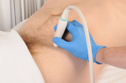
To perform ultrasound guided ilioinguinal nerve block, the inferior portion of linear high frequency ultrasound transducer is placed over the previously identified anterior superior iliac spine with the superior margin of the transducer pointed directly in an oblique plane at the ulbilicus.
From: https://www.oatext.com/A-simplified-approach-to-ultrasound-guided-ilioinguinal-nerve-block.php
Licencing
This work is licensed under a Creative Commons Attribution 4.0 International License.
File history
Click on a date/time to view the file as it appeared at that time.
| Date/Time | Thumbnail | Dimensions | User | Comment | |
|---|---|---|---|---|---|
| current | 20:40, 16 May 2023 |  | 412 × 274 (66 KB) | Jeremy (talk | contribs) | To perform ultrasound guided ilioinguinal nerve block, the inferior portion of linear high frequency ultrasound transducer is placed over the previously identified anterior superior iliac spine with the superior margin of the transducer pointed directly in an oblique plane at the ulbilicus. From: https://www.oatext.com/A-simplified-approach-to-ultrasound-guided-ilioinguinal-nerve-block.php |
You cannot overwrite this file.
File usage
The following page uses this file:


