File:Multifidus fat infiltration.png: Difference between revisions
From WikiMSK
(File uploaded with MsUpload) |
No edit summary |
||
| Line 1: | Line 1: | ||
Examples of amounts of fat in the lumbar multifidus muscles as seen on axial T1- weighted magnetic resonance imaging scans. | |||
These were rated as grade 0 if normal condition; grade 1 for slight fat infiltration (10–50%), and grade 2 for severe fat infiltration (>50%). | |||
Image and text are from an Open Access article distributed under the terms of the Creative Commons Attribution License (http://creativecommons.org/licenses/by/2.0), which permits unrestricted use, distribution, and reproduction in any medium, provided the original work is properly cited.<ref>{{#pmid:17254322}}</ref> | |||
Latest revision as of 14:56, 2 April 2021
Examples of amounts of fat in the lumbar multifidus muscles as seen on axial T1- weighted magnetic resonance imaging scans. These were rated as grade 0 if normal condition; grade 1 for slight fat infiltration (10–50%), and grade 2 for severe fat infiltration (>50%).
Image and text are from an Open Access article distributed under the terms of the Creative Commons Attribution License (http://creativecommons.org/licenses/by/2.0), which permits unrestricted use, distribution, and reproduction in any medium, provided the original work is properly cited.[1]
File history
Click on a date/time to view the file as it appeared at that time.
| Date/Time | Thumbnail | Dimensions | User | Comment | |
|---|---|---|---|---|---|
| current | 14:54, 2 April 2021 | 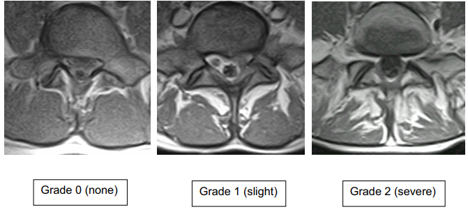 | 671 × 307 (191 KB) | Jeremy (talk | contribs) | File uploaded with MsUpload |
You cannot overwrite this file.
File usage
There are no pages that use this file.

