File:Neck tongue syndrome anatomy.jpg: Difference between revisions
(The relationship between the C2 dorsal root ganglion (g), spinal nerve (sn), and ventral ramus (vr, and the lateral atlanto-axial joint (j), posterior arch of the atlas (paa), the lamina of the axis (la), and the vertebral artery (va). A: Schematic illustrations of a dorsal view. B: Dissection of a right dorsolateral view. dr=C2 dorsal ramus (cut at its point of branching). cp=cervical plexus. ton=third occipital nerve. c=communicating branch C2 to C3. paam=posterior atlantoaxial membrane. C...) |
No edit summary |
||
| Line 6: | Line 6: | ||
Bogduk N. An anatomical basis for the neck-tongue syndrome. J Neurol Neurosurg Psychiatry. 1981 Mar;44(3):202-8. doi: 10.1136/jnnp.44.3.202. PMID: 7229642; PMCID: PMC490891. | Bogduk N. An anatomical basis for the neck-tongue syndrome. J Neurol Neurosurg Psychiatry. 1981 Mar;44(3):202-8. doi: 10.1136/jnnp.44.3.202. PMID: 7229642; PMCID: PMC490891. | ||
== Licencing == | == Licencing == | ||
{{Copyrighted}} | {{Copyrighted-permission|BMJ|26-3-22}} | ||
Latest revision as of 10:47, 26 March 2022
Summary
The relationship between the C2 dorsal root ganglion (g), spinal nerve (sn), and ventral ramus (vr, and the lateral atlanto-axial joint (j), posterior arch of the atlas (paa), the lamina of the axis (la), and the vertebral artery (va). A: Schematic illustrations of a dorsal view. B: Dissection of a right dorsolateral view. dr=C2 dorsal ramus (cut at its point of branching). cp=cervical plexus. ton=third occipital nerve. c=communicating branch C2 to C3. paam=posterior atlantoaxial membrane.
Copyright BMJ 1981. Used with permission Bogduk N. An anatomical basis for the neck-tongue syndrome. J Neurol Neurosurg Psychiatry. 1981 Mar;44(3):202-8. doi: 10.1136/jnnp.44.3.202. PMID: 7229642; PMCID: PMC490891.
Licencing
This file is copyrighted by BMJ, and is reproduced by permission obtained on 26-3-22.
File history
Click on a date/time to view the file as it appeared at that time.
| Date/Time | Thumbnail | Dimensions | User | Comment | |
|---|---|---|---|---|---|
| current | 10:46, 26 March 2022 | 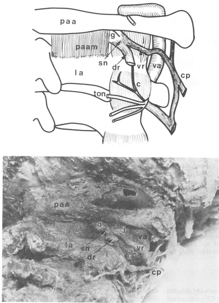 | 748 × 1,037 (203 KB) | Jeremy (talk | contribs) | The relationship between the C2 dorsal root ganglion (g), spinal nerve (sn), and ventral ramus (vr, and the lateral atlanto-axial joint (j), posterior arch of the atlas (paa), the lamina of the axis (la), and the vertebral artery (va). A: Schematic illustrations of a dorsal view. B: Dissection of a right dorsolateral view. dr=C2 dorsal ramus (cut at its point of branching). cp=cervical plexus. ton=third occipital nerve. c=communicating branch C2 to C3. paam=posterior atlantoaxial membrane. C... |
You cannot overwrite this file.
File usage
The following page uses this file:

