File:STT joint ultrasound tranducer placement smith.png: Difference between revisions
No edit summary |
No edit summary |
||
| Line 1: | Line 1: | ||
== Summary == | |||
The transducer is oriented nearly parallel to the first metacarpal and placed over the STT joint so that the joint appears in the centre of the display screen. The needle is advanced using an out-of-plane (short-axis) approach in a radial-to-ulnar direction and is advanced into the joint. | |||
== Licencing == | == Licencing == | ||
{{Copyrighted}} | {{Copyrighted}} | ||
{{#pmid:22039023}} | |||
©2011 by the American Institute of Ultrasound in Medicine | |||
Latest revision as of 16:17, 14 June 2021
Summary
The transducer is oriented nearly parallel to the first metacarpal and placed over the STT joint so that the joint appears in the centre of the display screen. The needle is advanced using an out-of-plane (short-axis) approach in a radial-to-ulnar direction and is advanced into the joint.
Licencing
![]() This file is copyrighted, and is reproduced in a limited way under the fair-use doctrine. It falls under the "Non-profit educational" clause of the Fair Use doctrine.
This file is copyrighted, and is reproduced in a limited way under the fair-use doctrine. It falls under the "Non-profit educational" clause of the Fair Use doctrine.
Smith et al.. Accuracy of sonographically guided and palpation guided scaphotrapeziotrapezoid joint injections. Journal of ultrasound in medicine : official journal of the American Institute of Ultrasound in Medicine 2011. 30:1509-15. PMID: 22039023. DOI.
©2011 by the American Institute of Ultrasound in Medicine
File history
Click on a date/time to view the file as it appeared at that time.
| Date/Time | Thumbnail | Dimensions | User | Comment | |
|---|---|---|---|---|---|
| current | 16:16, 14 June 2021 | 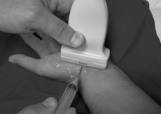 | 512 × 364 (133 KB) | Jeremy (talk | contribs) |
You cannot overwrite this file.
File usage
The following page uses this file:

