File:TMC joint ultrasound out of plane injection umphrey.png: Difference between revisions
From WikiMSK
No edit summary |
No edit summary |
||
| Line 1: | Line 1: | ||
==Summary== | |||
Ultrasound-guided injection of the TMC joint. A hypoechoic cleft is visualised between the trapezium (left) and first metacarpal (right) defining the joint space. The needle tip is visualised in the centre of the joint space. | |||
== Licencing == | == Licencing == | ||
{{Copyrighted}} | {{Copyrighted}} | ||
{{#pmid:18164346}} | |||
Latest revision as of 07:30, 15 June 2021
Summary
Ultrasound-guided injection of the TMC joint. A hypoechoic cleft is visualised between the trapezium (left) and first metacarpal (right) defining the joint space. The needle tip is visualised in the centre of the joint space.
Licencing
![]() This file is copyrighted, and is reproduced in a limited way under the fair-use doctrine. It falls under the "Non-profit educational" clause of the Fair Use doctrine.
This file is copyrighted, and is reproduced in a limited way under the fair-use doctrine. It falls under the "Non-profit educational" clause of the Fair Use doctrine.
Umphrey et al.. Ultrasound-guided intra-articular injection of the trapeziometacarpal joint: description of technique. Archives of physical medicine and rehabilitation 2008. 89:153-6. PMID: 18164346. DOI.
File history
Click on a date/time to view the file as it appeared at that time.
| Date/Time | Thumbnail | Dimensions | User | Comment | |
|---|---|---|---|---|---|
| current | 07:28, 15 June 2021 | 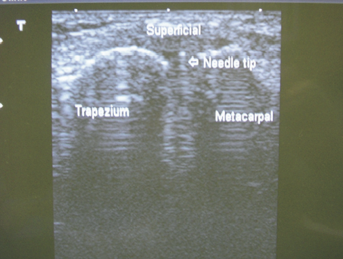 | 494 × 373 (324 KB) | Jeremy (talk | contribs) |
You cannot overwrite this file.
File usage
The following page uses this file:

