Finger Extensor Tendon Injuries: Difference between revisions
m (→Types) |
mNo edit summary |
||
| Line 21: | Line 21: | ||
== References == | == References == | ||
{{Article derivation|article-link=https://www.e-ultrasonography.org/journal/view.php?doi=10.14366/usg.15051|article=Current status of ultrasonography of the finger|author=Seun Ah Lee et al|license-link=https://creativecommons.org/licenses/by-nc/3.0/|license=CC-BY-NC}} | {{Article derivation|article-link=https://www.e-ultrasonography.org/journal/view.php?doi=10.14366/usg.15051|article=Current status of ultrasonography of the finger|author=Seun Ah Lee et al|license-link=https://creativecommons.org/licenses/by-nc/3.0/|license=CC-BY-NC}} | ||
[[Category:Hand and Wrist]] | [[Category:Hand and Wrist Conditions]] | ||
Revision as of 09:38, 3 March 2022
Extensor tendon tears typically occur in two locations in the finger.
Types
Mallet Finger
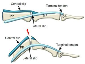
Mallet finger, which is commonly referred to as baseball finger or cricket finger, occurs at the distal phalangeal base, which is the insertion site of the extensor tendon, also known as the terminal tendon. This injury occurs when the distal interphalangeal (DIP) joint is abruptly forced into extreme flexion, at which time the tendon may pull off a piece of bone. The clinical symptoms of mallet finger include pain, swelling, and inability to extend the DIP joint
On US, instead of tendon echoes at the tendon insertion site of the distal phalanx, an irregular hypoechoic soft tissue lesion is usually present over the distal shaft of the middle phalanx, indicative of the retracted tendon end. In the case of an avulsion fracture, the detached bone fragment at the retracted tendon end and loss of substance in the distal phalangeal base can also be detected on US.
Boutonnière Deformity
The second most frequent site of injuries involving the extensor tendon is at or near the central slip insertion site of the middle phalangeal base. This injury is known as Boutonnière deformity. The mechanisms underlying Boutonnière deformity include acute violent flexion of the PIP joint, a blow to the dorsum of the middle phalanx, or volar dislocation of the PIP joint.
In the acute stage, Boutonnière deformity presents with pain, swelling of the PIP joint, mild extension lag, and reduced extension strength against resistance. After the acute stage, the intact lateral slips move the volar aspect, inducing flexion of the PIP joint, an increase in the force exerted on the intact terminal tendon insertion, and subsequent extension of the DIP joint.
On US, the central slip of the injured extensor tendon demonstrates a lack of tendon echoes that would indicate insertion into the middle phalangeal base, whereas the lateral slips are intact on both sides of the middle phalanx.
References
Part or all of this article or section is derived from Current status of ultrasonography of the finger by Seun Ah Lee et al, used under CC-BY-NC


