Ilioinguinal and Iliohypogastric Nerve Injection: Difference between revisions
(Created page with "{{procedure |indication= |syringe= |needle= |steroid= |local= |volume= }}The Ilioinguinal nerve injection, or nerve block, is a valuable diagnostic, prognostic, and therapeutic tool for managing groin and genital pain believed to be mediated via the ilioinguinal nerve ==Anatomy== The ilioinguinal nerve, comprised of fibers from the L1 nerve root with a contribution of fibers from the T12 nerve root in about 25% of patients, plays a critical role i...") |
No edit summary |
||
| Line 1: | Line 1: | ||
{{ | {{Authors}} | ||
{{Procedure | |||
| | |quality=Partial | ||
| | |indication=Chronic pelvic pain | ||
}} | |||
The Ilioinguinal nerve injection, or nerve block, is a valuable diagnostic, prognostic, and therapeutic tool for managing groin and genital pain believed to be mediated via the ilioinguinal nerve | |||
}}The Ilioinguinal nerve injection, or nerve block, is a valuable diagnostic, prognostic, and therapeutic tool for managing groin and genital pain believed to be mediated via the ilioinguinal nerve | |||
==Anatomy== | ==Anatomy== | ||
Revision as of 20:43, 16 May 2023
| Ilioinguinal and Iliohypogastric Nerve Injection | |
|---|---|
| Indication | Chronic pelvic pain |
The Ilioinguinal nerve injection, or nerve block, is a valuable diagnostic, prognostic, and therapeutic tool for managing groin and genital pain believed to be mediated via the ilioinguinal nerve
Anatomy
The ilioinguinal nerve, comprised of fibers from the L1 nerve root with a contribution of fibers from the T12 nerve root in about 25% of patients, plays a critical role in the sensation of the groin and genital regions. It follows a curvilinear course from the L1 and occasionally T12 somatic nerves, passing along the inside of the ilium's concavity. This nerve then continues to pass anteriorly within a fascial plane between the internal oblique and transverse abdominis muscles, where it is readily identified with ultrasound scanning.
At the anterior superior iliac spine level, the ilioinguinal nerve perforates anteriorly through the transverse abdominis muscle. Its terminal branches provide sensory innervation to the skin over the lower portion of the rectus abdominis muscle. Interconnections often exist between the ilioinguinal nerve and the adjacent iliohypogastric nerve, and occasionally the genitofemoral nerve.
Indications and Efficacy
Contraindications
Pre-procedural Evaluation
Equipment
Technique
Ultrasound Guided
- Patient Position: Supine
- Ultrasound Technique: Use a high frequency linear ultrasound transducer
- Start with the anterior superior iliac spine as the initial bony landmark
- Identify the umbilicus and palpate
- Place the inferior portion of the ultrasound transducer over the anterior superior iliac spine with the superior margin of the transducer pointed toward the umbilicus
- Rotate the superior margin of the ultrasound transducer superiorly and inferiorly until the fascial plane between the internal oblique and transverse abdominis muscle is identified
- Identify the ilioinguinal nerve within this fascial plane
- Identify the deep circumflex artery that lies within the same fascial plane adjacent to the target nerve.
- Needle Approach: From a point just below the inferior border of the ultrasound transducer, move the needle in-plane towards the ilioinguinal nerve
- Visualize needle tip and ilioinguinal nerve in real time
- Place needle tip within the fascial plane adjacent to the ilioinguinal nerve
- Needle Type: 1.5 inch (3.8 cm), 22-gauge needle
- Injection Solution: 5 mL of local anesthetic, and if necessary, 40-80 mg of methylprednisolone
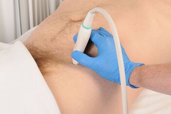
To perform ultrasound guided ilioinguinal nerve block, the inferior portion of linear high frequency ultrasound transducer is placed over the previously identified anterior superior iliac spine with the superior margin of the transducer pointed directly in an oblique plane at the ulbilicus.
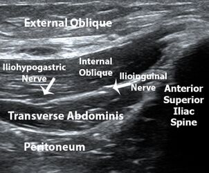
Oblique ultrasound image demonstrating the acoustic shadow of the anterior superior iliac spine and the muscles layers and facial plane containing the ilioinginal and iliohypogastric nerves.


Landmark Guided
- Patient Position: Supine
- Procedure:
- Identify the anterior superior iliac spine as the primary landmark
- Identify the umbilicus as the secondary landmark
- Prepare the skin overlying the identified landmarks with an antiseptic solution
- Insert the needle at a point just inferior to the anterior superior iliac spine, aiming towards the umbilicus
- Aspirate to ensure the needle is not in a blood vessel before injecting the anesthetic
- Needle Type: 1.5 inch (3.8 cm), 22-gauge needle
- Injection Solution: 5 mL of local anesthetic, and if necessary, 40-80 mg of methylprednisolone
Complications
The most feared complication of the anatomic landmark guided ilioinguinal nerve block is the inadvertent placement of the needle too deeply, leading to the needle tip entering the peritoneal cavity. Ultrasound guidance clearly delineates the margin between the transverse abdominis muscle and the underlying peritoneal cavity. In addition, the use of ultrasound can help to avoid inadvertent damage to the deep circumflex iliac artery, which lies within the same fascial plane and in close proximity to the ilioinguinal nerve.
Aftercare
Videos
See Also
External Links
Ultrasound-Guided Blocks for Pelvic Pain - NYSORA
Simplified ilioinguinal injection - OA Text
References
Literature Review
 Literature Review
- Reviews from the last 7 years: review articles, free review articles, systematic reviews, meta-analyses, NCBI Bookshelf
- Articles from all years: PubMed search, Google Scholar search.
- TRIP Database: clinical publications about evidence-based medicine.
- Other Wikis: Radiopaedia, Wikipedia Search, Wikipedia I Feel Lucky, Orthobullets,

