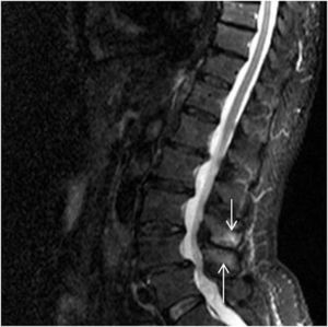
Interspinous Oedema
There are a variable number of bursae in the spinal column. They are normally located between the spinous processes of the cervical and lumbar segments. Bursitis of these structures can result in neck or back pain. In the neck, interspinous bursitis has been associated with several rheumatological conditions such as polymyalgia rheumatica, rheumatoid arthritis, and crystalopathies. In the lumbar spine the condition is also known as "kissing-spine" or Baastrup's disease and occurs in the context of degenerative change. An open access review of kissing spines is available by Filippiadis et al.[1]
Lumbar Kissing Spines

This condition, also known as Baastrup's disease, arises due to excessive lumbar lordosis, or following extension injuries to the lumbar spine. The adjacent spinous processes, the most common level being L4/5, compresses the intervening interspinous ligament. It is more common in the degenerative lumbar spine in those aged 70 and older, with no gender predilection, with changes occurring in the context of disc height loss, spondylolisthesis, and spondylosis.[1][2]
Aetiology
Bogduk theorises that the pain arises as a result of a periostitis of the spinous processes, or inflammation of the interspinous ligament. The periosteum of the spinous processes and the interspinous spaces are innervated by the medial branches of the lumbar dorsal rami. [2]
Clinical Features
Patients may have midline back pain that worsens with extension and relieved by flexion. On examination the patient may be tender over the suspected level. Rarely an epidural cystic mass may cause neurogenic claudication with extension.[1]
Imaging
Imaging includes radiography, CT, and MRI. The hallmark finding suggestive of kissing spines is close approximation and contact of adjacent spinous processes with all the subsequent findings, including oedema, cystic lesions, sclerosis, flattening and enlargement of the articulating surfaces, bursitis and occasionally epidural cysts or midline epidural fibrotic masses.[1]
On x-rays, the most common finding is the close approximation and contact of adjacent spinous processes. There may be sclerosis of the articulating surfaces. In more severe cases, there is flattening and enlargement of the articulating surfaces or articulation of the two affected spinous processes. Usually generalised degenerative spinal changes are seen, most prominent at the affected level.[1]
CT shows close approximation and contact of the adjacent spinous processes. There is sclerosis, flattening, and enlargement of the articulating surfaces or the articulation of the two affected spinous processes. In rare cases there may be soft tissue nodules on the sides of the spinous process, and this could represent dissection of the spinal bursa superficial to the erector spinae. There are usually findings of degenerative changes such as facet joint hypertrophy, disc herniation, and spondylolisthesis.[1]
On MRI, under 10% of symptomatic patients have findings of interspinous bursitis. Bursitis appears as a fluid-like signal located between the two suspect spinous processes. Other findings include flattening, sclerosis, enlargement, cystic lesions, and bone oedema affecting the articulating surfaces of the two spinous processes. There may be oedema of the interspinous ligament. Gadolinium administration may show enhancement of this area. Rarely there may be epidural cysts or epidural fibrotic masses causing dural compression.[1]
Treatment
Treatment options include NSAIDs, corticosteroid injections, and surgical therapies which include excision of the bursa or osteotomy. Injections, performed at the level of the interspinous ligament, should ideally be done with imaging guidance to ensure accurate needle positioning. Surgical therapy is not always successful. There may be additional factors in the patients pain such as facet joint irritation.
References
- ↑ 1.0 1.1 1.2 1.3 1.4 1.5 1.6 1.7 Filippiadis et al.. Baastrup's disease (kissing spines syndrome): a pictorial review. Insights into imaging 2015. 6:123-8. PMID: 25582088. DOI. Full Text.
- ↑ 2.0 2.1 Bogduk, Nikolai. Clinical and radiological anatomy of the lumbar spine. Chapter 15. Edinburgh: Elsevier/Churchill Livingstone, 2012.
Literature Review
- Reviews from the last 7 years: review articles, free review articles, systematic reviews, meta-analyses, NCBI Bookshelf
- Articles from all years: PubMed search, Google Scholar search.
- TRIP Database: clinical publications about evidence-based medicine.
- Other Wikis: Radiopaedia, Wikipedia Search, Wikipedia I Feel Lucky, Orthobullets,


