◔
Lumbar Spine Biomechanics
From WikiMSK
This article is a stub.
Movements
| Movement | Description | Movement occurs when | Force |
|---|---|---|---|
| Translation | Every point on a bone moving in the same direction and to the same extent | Single force or net single force acts on a bone | Shear force |
| Rotation | All the points on a bone move in parallel around a curved path that is centred on a fixed point, but to different extents depending on their distance from the centre of rotation. May be due to two opposed muscular actions, or muscular action and ligamentous resistance, or gravity opposed by either muscular action or ligamentous resistance. | Two unaligned forces acting in opposing directions on different parts of the bone forming a force couple | Torque |
- Axis of rotation
- Formal definition = that region that does not move when two or more opposing, unaligned forces act on a bone.
- With rotation of a bone, all the points on the bone can be grouped into individual planes that lie parallel to the direction of movement.
- In each plane, the points move about a centre located in that plane
- When all the centres of all the planes line up they depict a straight line forming the axis of rotation of the bone.
- It is a region where all opposing forces cancel out, and there is no net force acting, and so it stays stationary.
- Different forces create different axes of rotation.
Planes of Movement
Stress-Strain

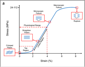
Typical stress–strain curve and schematization of the behavior of the collagen fibers for tendons. Typical ranges of stress and strain are indicated on the x and y axes.[1]

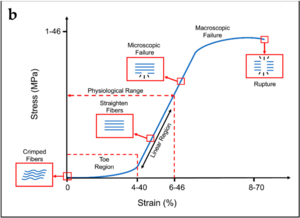
Typical stress–strain curve and schematization of the behavior of the collagen fibers for ligaments.[1]
- With stretching of a collagen fibre, the fibre resists elongation due to a resistance force from the chemical bonds between the collagen fibrils, between tropocollagen molecules, between collagen fibres, and between collagen fibres and proteoglycans.
- Stress = the applied elongating force, measured in units of force (newtons)
- Strain = the extent to which a fibre is elongated, measured as the fractional or percentage increase in length relative to the initial length. A fibre of length L0 when stretched to a new length L1 undergoes a strain of L1/L0 x 100%
- Tension strain = this occurs with a deforming force causing a structure to be stretched longitudinally
- Compression strain = this occurs with a deforming force causing a structure to be squashed. Measured as the fractional or percentage decrease in height of a structure
- Shear force = forces that cause two vertebrae to slides with respect to one another
- Shear strain = the strain that occurs in the intervening intervertebral disc
- Shear vs tension: tension conventionally applies to forces that are exerted along the long axis of a structure. Shear forces are applied across the axis.
- Torsion = when an object twists, the force is a torque, and the resultant strain is a torsion strain.
- Crimp = At rest, single collagen fibres are usually buckled, the wavy shape is called crimp
- Failure = Collagen fibres have broken and cease to resist elongation
Stress-strain curve
- Three main regions
- First region is the toe phase. This is where the crimp is being removed from the collagen fibre with the application of stress, this requires little energy due to no major chemical bonds
- The second, linear region is the steep slope along the middle of the curve, and is where the stress stretches the collagen fibre longitudinally and the crimp has been removed. The bonds within and between the collagen fibrils and tropocollagen molecules are being strained and some are being broken. The peak is the phase of failure of the collagen fibre, with substantial numbers of bonds being irreversibly broken.
- The last part of the curve is once failure has occurred, and elongation occurs with ever decreasing amounts of stress Collagen fibrils start to be strained and broken somewhere after 3% and 4% of elongation of the fibre. About 4% is the maximum a fibre can sustain without risking microscopic damage.
- Collagenous tissues, like ligaments and joint capsules, behave similarly to isolated collagen fibres and show similar stress-strain curves, but with additional mechanical events
- In the toe phase, in addition to the removal of crimp, this phase may also represent the removal of any macroscopic slack in the ligament
- In the second phase, the collagen fibres are being re-arranged in the stress structure. Fibres that are curved or run obliquely at rest are straightened to line up with the applied force. Water and proteoglycans may be displaced. Bonds between separate collagen fibres and between collagen fibres and their surrounding proteglycan matrix are strained. This results in a steep slope of the second phase. After re-arrangement of the fibres and displacement of water, then the individual collagen fibres are strained.
- It isn't known what proportion of collagen fibres need to fail before macroscopic failure occurs, and cannot be predicted, therefore different structures need to be separately tested with several samples to produce an average stress-strain curve of a specific structure.
- A very important point is omitted from Bogduk's lumbar spine anatomy book. Tendons have specific load transfer functions, and the toe region of their stress-strain curve is short (2-5%), and is similar with each tendon in the body. Ligaments on the other hand allow different ranges of motion in different joints, and have wider ranges of strain for the toe region depending on the anatomical site (ACL is 4%, spine ligaments are 10-40%).[1]
- Clinical examination only evaluates no further than just beyond the toe phase.
Stiffness
- Stiffness = resistance to deformation
- Measured by the force required to product a unit elongation or deformation
- The slope of the stress-strain curve of a structure
- Stiffer structures resist deformation and have a steeper stress-strain curve
- It implies greater degree of bonding between collagen fibres, or between collagen fibres and their surrounding matrix
Initial Range of Movement
Creep
Hysteresis
Fatigue Failure
Forces and Moments
Bibliography
- Bogduk, Nikolai. Clinical and radiological anatomy of the lumbar spine. Chapter 17. Edinburgh: Elsevier/Churchill Livingstone, 2012.
References
Literature Review
- Reviews from the last 7 years: review articles, free review articles, systematic reviews, meta-analyses, NCBI Bookshelf
- Articles from all years: PubMed search, Google Scholar search.
- TRIP Database: clinical publications about evidence-based medicine.
- Other Wikis: Radiopaedia, Wikipedia Search, Wikipedia I Feel Lucky, Orthobullets,


