Lumbar Spine Radiographs: Difference between revisions
From WikiMSK
No edit summary |
No edit summary |
||
| Line 5: | Line 5: | ||
==Lateral Views== | ==Lateral Views== | ||
[[File:Lumbar Spine Lateral Radiograph Normal.png|thumb|right|Lateral radiograph of the lumbar spine]] | |||
*Evident: Vertebral bodies and posterior elements | *Evident: Vertebral bodies and posterior elements | ||
*Trace the anatomy, start with L3 which is usually the least obscured then repeat with other vertebrae | *Trace the anatomy, start with L3 which is usually the least obscured then repeat with other vertebrae | ||
| Line 27: | Line 28: | ||
**Quadratus lumborum centrally, behind and lateral to the psoas | **Quadratus lumborum centrally, behind and lateral to the psoas | ||
**Lumbar and lower thoracic erector spinae posteriorly, lateral to the multifidus, fibres running cephalad and ventrally. | **Lumbar and lower thoracic erector spinae posteriorly, lateral to the multifidus, fibres running cephalad and ventrally. | ||
<gallery widths=400px heights=350px> | |||
Lumbar Spine L3 Lateral Normal.png|Lateral radiograph of L3 | |||
Lumbar Spine L4-5 Lateral Normal.png|Lateral radiograph the L4-5 zygapophyseal joint | |||
</gallery> | |||
==Anterior (or Posterior) View== | |||
[[File:Lumbar Spine AP Radiograph Normal.png|thumb|right|AP radiograph of the lumbar spine]] | |||
==References== | ==References== | ||
*Bogduk, Nikolai. Clinical and radiological anatomy of the lumbar spine. Chapter 18. Edinburgh: Elsevier/Churchill Livingstone, 2012. | *Bogduk, Nikolai. Clinical and radiological anatomy of the lumbar spine. Chapter 18. Edinburgh: Elsevier/Churchill Livingstone, 2012. | ||
[[Category:Lumbar Spine]] | [[Category:Lumbar Spine]] | ||
Revision as of 14:20, 3 May 2021
- Bones are transparent and so where bony structures overlap this complicates interpretation of plan x-rays of the lumbar spine.
- Lateral x-rays have few superimposed structures
- Anteroposterior views have multiple superimposed elements.
- Interpretation is through "anatomy by expectation" - expect what should be there, and then decide if what they expect is indeed present.
Lateral Views

- Evident: Vertebral bodies and posterior elements
- Trace the anatomy, start with L3 which is usually the least obscured then repeat with other vertebrae
- Vertebral body: Superior, anterior, and inferior margins. L5 has a large base that flows onto the pedicle and vertebral body of L5
- Pedicle: at the posterior margin of the vertebral body
- Superior articular process: starts on the posterior superior corner of the pedicle, projects dorsally and cephalad, with a rounded mushroom shaped head
- Inferior articular process: From the posterior inferior corner of the pedicle, narrow lamina projects caudally and slightly dorsally, and expands into a rounded mass.
- Transverse process: At the junction between the pedicle and the superior articular process, elliptical shadow
- Spinous process: profile of the blade of an axe, arises from the back of the lamina
- Interpretation of the facet joints
- The superior articular processes cover the inferior articular processes of the vertebra above.
- Multiple markings can appear in the facet joints, creating the illusion that the joint space projects laterally.
- This occurs when C-shaped or J-shaped joints are viewed from the side.
- The joint space seen is only the ventral aspect of the joint, the rest of the joint projects laterally and is covered by the superior articular process.
- Soft tissues to expect but can't see
- Dural sac behind the vertebral bodies
- Spinal nerves in the intervertebral foramina
- Psoas major clamping the lumbar arteries and lumbar veins against the vertebral bodies
- Right crus and IVC anteriorly towards the right
- Aorta and left crus anteriorly towards the left
- Multifidus posteriorly behind the laminae and against the spinous process, fibres passing dorsally and cephalad
- Quadratus lumborum centrally, behind and lateral to the psoas
- Lumbar and lower thoracic erector spinae posteriorly, lateral to the multifidus, fibres running cephalad and ventrally.
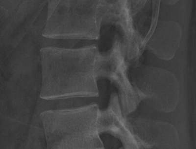
Lateral radiograph of L3
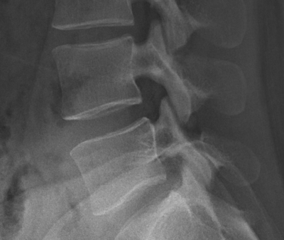
Lateral radiograph the L4-5 zygapophyseal joint


Anterior (or Posterior) View

References
- Bogduk, Nikolai. Clinical and radiological anatomy of the lumbar spine. Chapter 18. Edinburgh: Elsevier/Churchill Livingstone, 2012.

