Lumbar Spine Radiographs
Plan x-rays of the lumbar spine provide little diagnostic value compared to CT and MRI. However the skill is useful in fluoroscopy and much of the knowledge and practice is transferrable to interpreting more advanced imaging. Interpretation can be complex because of the transparency of bones leading to overlapping structures especially in the anteroposterior view. Interpretation is through "anatomy by expectation" - expect what should be there, and then decide if what they expect is indeed present, while ignoring distracting lines.
Lateral Views
Interpretation

- Evident: Vertebral bodies and posterior elements
- Trace the anatomy, start with L3 which is usually the least obscured then repeat with other vertebrae
- Vertebral body: Superior, anterior, and inferior margins. L5 has a large base that flows onto the pedicle and vertebral body of L5
- Pedicle: at the posterior margin of the vertebral body
- Superior articular process: starts on the posterior superior corner of the pedicle, projects dorsally and cephalad, with a rounded mushroom shaped head
- Inferior articular process: From the posterior inferior corner of the pedicle, narrow lamina projects caudally and slightly dorsally, and expands into a rounded mass.
- Transverse process: At the junction between the pedicle and the superior articular process, elliptical shadow
- Spinous process: profile of the blade of an axe, arises from the back of the lamina
- Interpretation of the facet joints
- The superior articular processes cover the inferior articular processes of the vertebra above.
- Multiple markings can appear in the facet joints, creating the illusion that the joint space projects laterally.
- This occurs when C-shaped or J-shaped joints are viewed from the side.
- The joint space seen is only the ventral aspect of the joint, the rest of the joint projects laterally and is covered by the superior articular process.
- Soft tissues to expect but can't see
- Dural sac behind the vertebral bodies
- Spinal nerves in the intervertebral foramina
- Psoas major clamping the lumbar arteries and lumbar veins against the vertebral bodies
- Right crus and IVC anteriorly towards the right
- Aorta and left crus anteriorly towards the left
- Multifidus posteriorly behind the laminae and against the spinous process, fibres passing dorsally and cephalad
- Quadratus lumborum centrally, behind and lateral to the psoas
- Lumbar and lower thoracic erector spinae posteriorly, lateral to the multifidus, fibres running cephalad and ventrally.
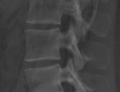
Zoomed in lateral view of L3
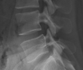
Zoomed in lateral view of L4-5 zygapophyseal joint


Anterior (or Posterior) View
AP is usually done, although PA dramatically reduces the effective radiation exposure by 41% without any effect on the image quality.[1]
Interpretation


- Superimposition makes AP views complex to interpret due to the lordosis and superimposition of the posterior elements on the anterior elements
- Upper vertebrae are tilted cephalad, and lower vertebrae are tilted caudad
- middle vertebral body margins are horizontal, while upper and lower are elliptical.
- In upper bodies, the cephalad margin is the anterior margin of the body, and caudad margin is the posterior margin
- In lower bodies, the cephalad margin is the posterior margin of the body, and caudad margin is the anterior margin.
- Regions
- Vertebral body
- Rectangular shadow with a scalloped waist
- Scalloped lateral margin runs tangential or near to the lateral margin of the pedicle
- Superior surface ellipses may appear like muffin tops
- L5 may be difficult to discern because of severe lordotic tilt, so that the vertebral body is below most of the posterior elements, and also because it is wedge shaped.
- Pedicles:
- Pair of vertical pillars, described as 'eyes.'
- Transverse processes
- Project from the pedicles.
- May be hard to visualise due to being thin
- L5 may be narrower than expected due to steep tilting
- Inferior margin flows into the lateral margin of the lamina., opposite the inferolateral corner of the pedicle.
- Laminae
- Rectangular plates
- Superior margin clearly evident towards the midline
- Laterally the superior margin is obscured by the inferior articular process, and can't see well the continuity with the superior articular process.
- Facet joints
- Where joint spaces and articular margins are present, straight vertical lines correspond to the articular surfaces, with sharp corners.
- Where joint spaces and articular margins aren't present, the joints face coronally, with overlapping superior and inferior articular processes, with rounded corners.
- Curved surfaces of the superior and inferior articular processes
- Superior articular processes: convex outer margin curves medially and inferiorly, across the pedicle, becoming continuous with the lateral margin of the lamina.
- Inferior articular processes: curvature continues upwards to become the inferior margin of the lamina.
- Superior and inferior articular processes: project from the corners of the laminae
- Vertebral body
- Soft tissues to expect but can't see
- Anteriorly
- Left and right cruri at the upper lumbar levels
- Lumbar sympathetic trunks throughout the length of the vertebral column
- Quadratus lumborum lateral to the column
- Psoas major covers the column
- Inferior vena cava covers the vertebral bodies as far as L5
- Aorta covers the vertebral bodies as far as L4
- Posteriorly
- Multifidus covers the laminae
- Longissimus thoracis pars lumborum aims for the accessory processes
- Iliocostalis lumborum pars lumborum aims for the tips of the tranverse processes
- Anteriorly
Additional Projections
Oblique view

- Used to visualise the articular facets and pars interarticularis of the lumbar spine
- Also used for needle placement in transforaminal epidural corticosteroid injections
- Scottie dog isnormal appearance of the lumbar spine when seen on oblique radiographic projection
- On oblique views, the posterior elements of the vertebra form the figure of a Scottie dog with
- the transverse process being the nose
- the pedicle forming the eye
- the inferior articular facet being the front leg
- the superior articular facet representing the ear
- the pars interarticularis (the portion of the lamina that lies between the facets) equivalent to the neck of the dog.
- If spondylolysis is present, the pars interarticularis, or the neck of the dog, will have a defect or break. It often looks as if the dog has a collar around the neck (or decapitation for those with a bloodier imagination).[2]
Flexion-extension view
- functional view used to assess spinal stability
Bibliography
- Bogduk, Nikolai. Clinical and radiological anatomy of the lumbar spine. Chapter 18. Edinburgh: Elsevier/Churchill Livingstone, 2012.
References
- ↑ Green et al.. Lumbar spine radiographs - is it time for widespread adoption of posteroanterior projection?. The British journal of radiology 2019. 92:20190386. PMID: 31356113. DOI. Full Text.
- ↑ Assoc Prof Craig Hacking and Assoc Prof Frank Gaillard et al. Scottie dog sign (spine). https://radiopaedia.org/articles/scottie-dog-sign-spine

