◒
Lumbar Spine Radiographs
From WikiMSK
This article is still missing information.
Plan x-rays of the lumbar spine provide little diagnostic value compared to CT and MRI. However the skill is useful in fluoroscopy and much of the knowledge and practice is transferrable to interpreting more advanced imaging. Interpretation can be complex because of the transparency of bones leading to overlapping structures especially in the anteroposterior view. Interpretation is through "anatomy by expectation" - expect what should be there, and then decide if what they expect is indeed present, while ignoring distracting lines.
Lateral Views

- Evident: Vertebral bodies and posterior elements
- Trace the anatomy, start with L3 which is usually the least obscured then repeat with other vertebrae
- Vertebral body: Superior, anterior, and inferior margins. L5 has a large base that flows onto the pedicle and vertebral body of L5
- Pedicle: at the posterior margin of the vertebral body
- Superior articular process: starts on the posterior superior corner of the pedicle, projects dorsally and cephalad, with a rounded mushroom shaped head
- Inferior articular process: From the posterior inferior corner of the pedicle, narrow lamina projects caudally and slightly dorsally, and expands into a rounded mass.
- Transverse process: At the junction between the pedicle and the superior articular process, elliptical shadow
- Spinous process: profile of the blade of an axe, arises from the back of the lamina
- Interpretation of the facet joints
- The superior articular processes cover the inferior articular processes of the vertebra above.
- Multiple markings can appear in the facet joints, creating the illusion that the joint space projects laterally.
- This occurs when C-shaped or J-shaped joints are viewed from the side.
- The joint space seen is only the ventral aspect of the joint, the rest of the joint projects laterally and is covered by the superior articular process.
- Soft tissues to expect but can't see
- Dural sac behind the vertebral bodies
- Spinal nerves in the intervertebral foramina
- Psoas major clamping the lumbar arteries and lumbar veins against the vertebral bodies
- Right crus and IVC anteriorly towards the right
- Aorta and left crus anteriorly towards the left
- Multifidus posteriorly behind the laminae and against the spinous process, fibres passing dorsally and cephalad
- Quadratus lumborum centrally, behind and lateral to the psoas
- Lumbar and lower thoracic erector spinae posteriorly, lateral to the multifidus, fibres running cephalad and ventrally.
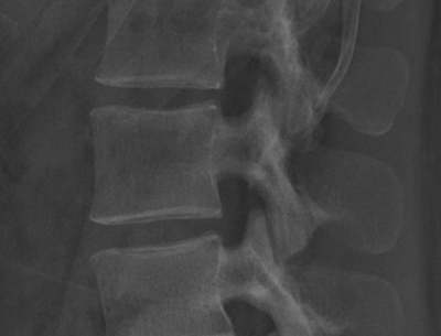
Zoomed in lateral view of L3
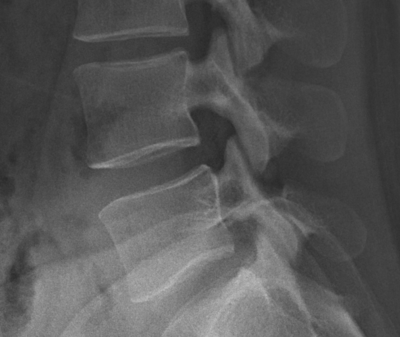
Zoomed in lateral view of L4-5 zygapophyseal joint


Anterior (or Posterior) View


- Superimposition makes AP views complex to interpret due to the lordosis and superimposition of the posterior elements on the anterior elements
- Upper vertebrae are tilted cephalad, and lower vertebrae are tilted caudad
- middle vertebral body margins are horizontal, while upper and lower are elliptical.
- In upper bodies, the cephalad margin is the anterior margin of the body, and caudad margin is the posterior margin
- In lower bodies, the cephalad margin is the posterior margin of the body, and caudad margin is the anterior margin.
References
- Bogduk, Nikolai. Clinical and radiological anatomy of the lumbar spine. Chapter 18. Edinburgh: Elsevier/Churchill Livingstone, 2012.

