Metatarsophalangeal Joints: Difference between revisions
No edit summary |
No edit summary |
||
| (3 intermediate revisions by the same user not shown) | |||
| Line 1: | Line 1: | ||
{{ | {{Joint | ||
|image= | |quality=Partial | ||
|caption= | |image=Metatarsophalangeal joints with latin names.png | ||
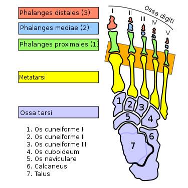
| | |caption=Bones of the foot labelled in latin with the MTPJs outlined in orange. | ||
|type= | |type=Synovial Joint | ||
|secondary-type=Condyloid Joint | |||
|bones=Metacarpal, Proximal Phalanx (Foot) | |bones=Metacarpal, Proximal Phalanx (Foot) | ||
|ligaments=Capsule, collateral ligamentous complexes, plantar plate. For great toe also sesamoid-phalangeal ligaments. | |ligaments=Capsule, collateral ligamentous complexes, plantar plate. For great toe also sesamoid-phalangeal ligaments. | ||
| Line 9: | Line 10: | ||
|innervation=Digital nerves from the ulnar and median nerves | |innervation=Digital nerves from the ulnar and median nerves | ||
|vasculature=Deep digital arteries from the superficial palmar arch | |vasculature=Deep digital arteries from the superficial palmar arch | ||
|conditions=[[Plantar Plate Injury (Turf Toe)|Turf Toe]], [[Plantar Plate Tears]],[[Freiberg Disease]] | |conditions=[[Plantar Plate Injury (Turf Toe)|Turf Toe]], [[Plantar Plate Tears]],[[Freiberg Disease]] | ||
}} | }} | ||
The five metatarsophalangeal joints are each composed of a convex metatarsal head and a concave proximal phalanx. | The five metatarsophalangeal joints are each composed of a convex metatarsal head and a concave proximal phalanx. | ||
== Motion == | ==Motion== | ||
The primary motion is dorsiflexion and plantarflexion, with a lesser amount of abduction and adduction. Passive range of motion is 65° for dorsiflexion, and 40° for plantarflexion for joints 2-5. The first MTPJ has 85° of dorsiflexion | The primary motion is dorsiflexion and plantarflexion, with a lesser amount of abduction and adduction. Passive range of motion is 65° for dorsiflexion, and 40° for plantarflexion for joints 2-5. The first MTPJ has 85° of dorsiflexion | ||
| Line 25: | Line 23: | ||
The windlass mechanism of the plantar aponeurosis provides stability to the medial aspect of the foot with great toe dorsiflexion. The peroneus longus muscle acts to press the metatarsal head into the ground when the body passes over the foot in toe-off. | The windlass mechanism of the plantar aponeurosis provides stability to the medial aspect of the foot with great toe dorsiflexion. The peroneus longus muscle acts to press the metatarsal head into the ground when the body passes over the foot in toe-off. | ||
== Ligaments == | ==Ligaments== | ||
{{Main|Ligaments of the Foot and Ankle}} | {{Main|Ligaments of the Foot and Ankle}} | ||
* Medial and lateral collateral ligaments: primary ligaments. | *Medial and lateral collateral ligaments: primary ligaments. | ||
* Four transverse metatarsal ligaments: bind the metatarsal heads together, providing stability for the forefoot. | *Four transverse metatarsal ligaments: bind the metatarsal heads together, providing stability for the forefoot. | ||
== Clinical Applications == | ==Clinical Applications== | ||
Narrow high-heeled shoe wearing can lead to mechanical entrapment of the interdigital nerves, most commonly the third. The nerves are pressed against the transverse intermetatarsal ligaments and metatarsal heads, leading to neuroma formation | Narrow high-heeled shoe wearing can lead to mechanical entrapment of the interdigital nerves, most commonly the third. The nerves are pressed against the transverse intermetatarsal ligaments and metatarsal heads, leading to neuroma formation | ||
First MTPJ stress fractures or inflammation can occur with excessive stress on the joint. Pain can occur in the sesamoid bones which are found within the flexor hallucis brevis tendon. | First MTPJ stress fractures or inflammation can occur with excessive stress on the joint. Pain can occur in the sesamoid bones which are found within the flexor hallucis brevis tendon. | ||
== References == | ==References== | ||
* Basic Biomechanics of the Musculoskeletal System - Nordin 4th edition 2012 | *Basic Biomechanics of the Musculoskeletal System - Nordin 4th edition 2012 | ||
[[Category:Foot and Ankle Anatomy]] | [[Category:Foot and Ankle Anatomy]] | ||
[[Category:Joints]] | [[Category:Joints]] | ||
[[Category:Joints of the Lower Limb]] | |||
Latest revision as of 16:55, 30 April 2022

| |
| Metatarsophalangeal Joints | |
|---|---|
| Primary Type | Synovial Joint |
| Secondary Type | Condyloid Joint |
| Bones | Metacarpal, Proximal Phalanx (Foot) |
| Ligaments | Capsule, collateral ligamentous complexes, plantar plate. For great toe also sesamoid-phalangeal ligaments. |
| Muscles | flexor digitorum longus, flexor digitorum brevis, extensor digitorum longus, extensor digitorum brevis, flexor digiti minimi brevis, abductor digiti minimi, dorsal and plantar Interossei, lumbricals |
| Innervation | Digital nerves from the ulnar and median nerves |
| Vasculature | Deep digital arteries from the superficial palmar arch |
| ROM | |
| Volume | |
| Conditions | Turf Toe, Plantar Plate Tears,Freiberg Disease |
The five metatarsophalangeal joints are each composed of a convex metatarsal head and a concave proximal phalanx.
Motion
The primary motion is dorsiflexion and plantarflexion, with a lesser amount of abduction and adduction. Passive range of motion is 65° for dorsiflexion, and 40° for plantarflexion for joints 2-5. The first MTPJ has 85° of dorsiflexion
During normal toe-off, the MTPJ needs to dorsiflex to 60°. Other tasks such as sitting on the heels or standing on tiptoes requires greater dorsiflexion.
There is some tangential sliding of the first MTPJ from maximum plantarflexion to moderate dorsiflexion, along with some compression dorsally at maximum dorsiflexion. With full dorsiflexion the metatarsal head surface contact area shifts dorsally as joint compression occurs. This is the reason for dorsal osteophyte formation and limited dorsiflexion of the proximal phalanx in hallux rigidus.
The windlass mechanism of the plantar aponeurosis provides stability to the medial aspect of the foot with great toe dorsiflexion. The peroneus longus muscle acts to press the metatarsal head into the ground when the body passes over the foot in toe-off.
Ligaments
- Main article: Ligaments of the Foot and Ankle
- Medial and lateral collateral ligaments: primary ligaments.
- Four transverse metatarsal ligaments: bind the metatarsal heads together, providing stability for the forefoot.
Clinical Applications
Narrow high-heeled shoe wearing can lead to mechanical entrapment of the interdigital nerves, most commonly the third. The nerves are pressed against the transverse intermetatarsal ligaments and metatarsal heads, leading to neuroma formation
First MTPJ stress fractures or inflammation can occur with excessive stress on the joint. Pain can occur in the sesamoid bones which are found within the flexor hallucis brevis tendon.
References
- Basic Biomechanics of the Musculoskeletal System - Nordin 4th edition 2012

