Peroneal Tendon Sheath Injection: Difference between revisions
No edit summary |
No edit summary |
||
| Line 2: | Line 2: | ||
{{procedure | {{procedure | ||
|image= | |image= | ||
|indication=[[Peroneal Tendinopathy]] | |indication=[[Peroneal Tendon Disorders|Peroneal Tendinopathy]] | ||
|syringe=3mL | |syringe=3mL | ||
|needle=25-gauge 1.5-inch needle | |needle=25-gauge 1.5-inch needle | ||
| Line 12: | Line 12: | ||
==Anatomy== | ==Anatomy== | ||
The peroneal tendons are located in the lateral compartment of the leg. They are the primary structures provided foot eversion. They are important active stabilisers of the ankle joint. Three are three categories of painful peroneal tendon disorders: tendinopathy, instability (subluxation and dislocation), and tears and ruptures. | |||
==Indications and Efficacy== | ==Indications and Efficacy== | ||
In a study of 96 patients who had peroneal tendon sheath injections, 25% progressed to surgery. 43.7% reported 0-1 weeks of pain relief, 12.6% 2-6 weeks, 6.9% 7-12 weeks, and 36.8% greater than 12 weeks. Preinjection duration of symptoms was associated with post-injection duration of pain relief (P=.036). Patients with less than 1 year of symptoms had a median 6-12 weeks of postinjection pain relief.{{#pmid:31068007|fram}} {{#pmid:31068007|fram}} | In a study of 96 patients who had peroneal tendon sheath injections, 25% progressed to surgery. 43.7% reported 0-1 weeks of pain relief, 12.6% 2-6 weeks, 6.9% 7-12 weeks, and 36.8% greater than 12 weeks. Preinjection duration of symptoms was associated with post-injection duration of pain relief (P=.036). Patients with less than 1 year of symptoms had a median 6-12 weeks of postinjection pain relief.{{#pmid:31068007|fram}} {{#pmid:31068007|fram}} | ||
| Line 27: | Line 28: | ||
*Visualise tendons running together at the level of the distal fibula | *Visualise tendons running together at the level of the distal fibula | ||
*Visualise needle during injection to confirm intrasheath, extratendinous injection. | *Visualise needle during injection to confirm intrasheath, extratendinous injection. | ||
===Landmark Guided=== | ===Landmark Guided=== | ||
Landmark guidance is only 60% accurate, vs 100% accurate for ultrasound. | Landmark guidance is only 60% accurate, vs 100% accurate for ultrasound. | ||
Revision as of 15:11, 2 June 2021
| Peroneal Tendon Sheath Injection | |
|---|---|
| Indication | Peroneal Tendinopathy |
| Syringe | 3mL |
| Needle | 25-gauge 1.5-inch needle |
| Steroid | 1mL of 40mg triamcinolone. |
| Local | 1mL of 1% lidocaine |
| Volume | 2mL |
Anatomy
The peroneal tendons are located in the lateral compartment of the leg. They are the primary structures provided foot eversion. They are important active stabilisers of the ankle joint. Three are three categories of painful peroneal tendon disorders: tendinopathy, instability (subluxation and dislocation), and tears and ruptures.
Indications and Efficacy
In a study of 96 patients who had peroneal tendon sheath injections, 25% progressed to surgery. 43.7% reported 0-1 weeks of pain relief, 12.6% 2-6 weeks, 6.9% 7-12 weeks, and 36.8% greater than 12 weeks. Preinjection duration of symptoms was associated with post-injection duration of pain relief (P=.036). Patients with less than 1 year of symptoms had a median 6-12 weeks of postinjection pain relief.[1] [1]
Contraindications
Pre-procedural Evaluation
Equipment
Technique
Ultrasound Guided
- Position: Lateral decubitus
- Outline distal fibula with a pen
- Clean area with alcohol, and load 1mL of 1% lidocaine and 1mL of 40mg triamcinolone.
- 25-gauge 1.5-inch needle
- High frequency probe in transverse position
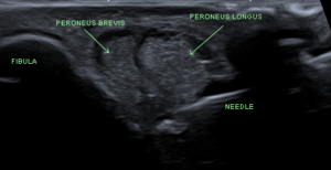
- Visualise tendons running together at the level of the distal fibula
- Visualise needle during injection to confirm intrasheath, extratendinous injection.
Landmark Guided
Landmark guidance is only 60% accurate, vs 100% accurate for ultrasound.
Complications
In a study of 96 patients, there were 2 reported complications (1.8%): 1 case of self-limited sural nerve irritation and 1 of peroneus longus tear progression.[1]
Aftercare
Videos
See Also
External Links
References
Literature Review
- Reviews from the last 7 years: review articles, free review articles, systematic reviews, meta-analyses, NCBI Bookshelf
- Articles from all years: PubMed search, Google Scholar search.
- TRIP Database: clinical publications about evidence-based medicine.
- Other Wikis: Radiopaedia, Wikipedia Search, Wikipedia I Feel Lucky, Orthobullets,


