◔
Peroneal Tendon Sheath Injection
From WikiMSK
This article is a stub.
| Peroneal Tendon Sheath Injection | |
|---|---|
| Indication | |
Anatomy
Indications and Efficacy
In a study of 96 patients who had peroneal tendon sheath injections, 25% progressed to surgery. 43.7% reported 0-1 weeks of pain relief, 12.6% 2-6 weeks, 6.9% 7-12 weeks, and 36.8% greater than 12 weeks. Preinjection duration of symptoms was associated with post-injection duration of pain relief (P=.036). Patients with less than 1 year of symptoms had a median 6-12 weeks of postinjection pain relief.[1] [1]
Contraindications
Pre-procedural Evaluation
Equipment
Technique
Ultrasound Guided
- Position: Lateral decubitus
- Outline distal fibula with a pen
- Clean area with alcohol, and load 1mL of 1% lidocaine and 1mL of 40mg triamcinolone.
- 25-gauge 1.5-inch needle
- High frequency probe in transverse position
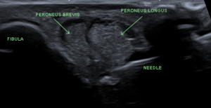
- Visualise tendons running together at the level of the distal fibula
- Visualise needle during injection to confirm intrasheath, extratendinous injection.
Fluoroscopy Guided
Landmark Guided
Landmark guidance is only 60% accurate, vs 100% accurate for ultrasound.
Complications
In a study of 96 patients, there were 2 reported complications (1.8%): 1 case of self-limited sural nerve irritation and 1 of peroneus longus tear progression.[1]
Aftercare
Videos
See Also
External Links
References
Literature Review
- Reviews from the last 7 years: review articles, free review articles, systematic reviews, meta-analyses, NCBI Bookshelf
- Articles from all years: PubMed search, Google Scholar search.
- TRIP Database: clinical publications about evidence-based medicine.
- Other Wikis: Radiopaedia, Wikipedia Search, Wikipedia I Feel Lucky, Orthobullets,


