Radiation Safety
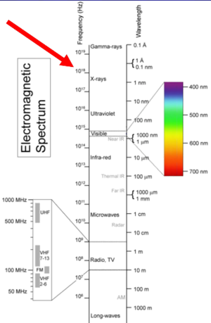
X-rays are on the electromagnetic spectrum, they are massless particles that travel at the speed of light. They have a very short wavelength which gives it certain properties. You can't see it, it passes through things, and it ionizes all matter. In a biological system it can give rise to unwanted effects. The x-ray scatter is very similar to visible light scatter. Think of x-rays of short wavelength light that is ionizing.
Every x-ray machine uses the same fundamental method of producing x-rays. It is measured in volts (kVs), and milliamps. The focal spot is very small - generated from a point. The intensity drops off over the square of the distance (1 / r^2). This is called the inverse square law related to divergence. Getting closer to the source causing a rapid rise in intensity. X-rays interact with matter via ionisation, and the intensity reduces exponentially reduces due to attenuation. These two phenomenon - divergence and attenuation - can be used to improve safety.
X-ray beams can be measured. Exposure is the easiest thing to measure and is the charge per mass in air. Fluoroscopy systems have an air chamber which collects the amount of charge in that small volume of air, and the ionization of air is measured. The absorbed dose related to the energy per mass. It is measured in Joules per kilogram in Grays (Gy). Systems will measure this in milli-grays. An orthopaedic type procedure may be around 100mGy, while a radiotherapy dose may be around 50Gy. The equivalent dose is the biologically weighted absorbed dose and is measured in Sievert (Sv). This relates to the overall risk to the patient. For x-rays 1 Gy = 1 Sv (whole body exposure). Ethics risk calculations and occupational exposure calculations use Sieverts.
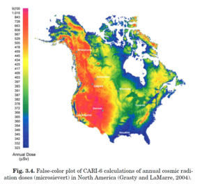
Natural background sources give around 2mSv. There is cosmic radiation, internal radiation, radon, mildly radioactive food. There is a slight variation in altitude with higher doses at higher altitudes. A patient dose is typically in 0.05-20mSv. Long cases may get up to 20mSv. Multi-phase CT scans can also get up to 20mSv. One of the challenges is that in over 100mSv as a single dose there is an increased risk of cancers. We live in this uncertain zone where we are working in a low level where we are not completely sure what the risk is.
Occupational exposure is similar to moving from a low altitude to a higher altitude area. Reducing population dose is focused on diagnostic procedures.
The total energy absorbed from x-rays) even at LD50 is very small. It is the microscopic energy deposition that is important (DNA the principal target). The repair of damage is very sophisticated (background radiation). Total repair leads to no effects. While mis-repaired or unrepaired damage leads to cell death (deterministic effects), or the cell can remain viable but have stochastic effects (cancer for somatic cells, and hereditary effects on germ cells)


