File list
From WikiMSK
This special page shows all uploaded files.
| Date | Name | Thumbnail | Size | User | Description | Versions |
|---|---|---|---|---|---|---|
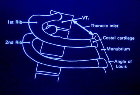
| 21:51, 18 February 2022 | Upper thoracic aperture.jpg (file) |  |
30 KB | Jeremy | Unknown source | 1 |
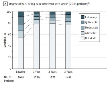
| 20:58, 17 February 2022 | Back and leg pain before and after bariatric surgery.jpg (file) |  |
24 KB | Jeremy | Degree of back or leg pain interfered with work before and after bariatric surgery. From King WC, Chen JY, Belle SH, Courcoulas AP, Dakin GF, Elder KA, Flum DR, Hinojosa MW, Mitchell JE, Pories WJ, Wolfe BM, Yanovski SZ. Change in Pain and Physical Function Following Bariatric Surgery for Severe Obesity. JAMA. 2016 Apr 5;315(13):1362-71. doi: 10.1001/jama.2016.3010. PMID: 27046364; PMCID: PMC4856477. | 1 |
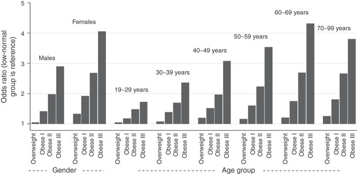
| 20:39, 17 February 2022 | Obesity pain odds ratio.jpg (file) |  |
38 KB | Jeremy | Odds ratios for pain yesterday for BMI classifications by gender and age groups, where the Low-Normal BMI group is the reference group. From Stone AA, Broderick JE. Obesity and pain are associated in the United States. Obesity (Silver Spring). 2012 Jul;20(7):1491-5. doi: 10.1038/oby.2011.397. Epub 2012 Jan 19. Erratum in: Obesity (Silver Spring). 2012 Jul;20(7):1546. PMID: 22262163. | 1 |
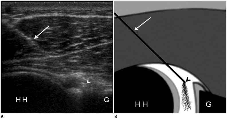
| 20:25, 14 February 2022 | GHJ injection under US.jpg (file) |  |
41 KB | Jeremy | Ultrasonography-guided posterior injection technique for shoulder MR arthrography. A. Transverse ultrasonography image shows needle track and intraarticular needle tip in patient with posterior glenohumeral joint puncture for MR arthrography. It also reveals distention of joint capsule with free fluid within joint space. White arrow = needle shaft, white arrowhead = intraarticular needle tip. B. Corresponding schematic drawing shows optimal needle track and placement. White arrow = needle sha... | 1 |
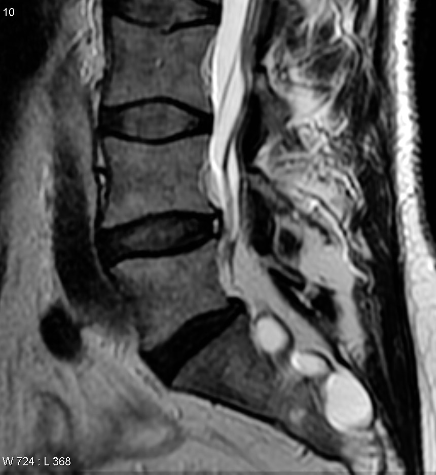
| 19:52, 14 February 2022 | Tarlov cyst and annular fissure.jpg (file) |  |
106 KB | Jeremy | Case courtesy of Assoc Prof Frank Gaillard, <a href="https://radiopaedia.org/?lang=gb">Radiopaedia.org</a>. From the case <a href="https://radiopaedia.org/cases/12574?lang=gb">rID: 12574</a> | 1 |
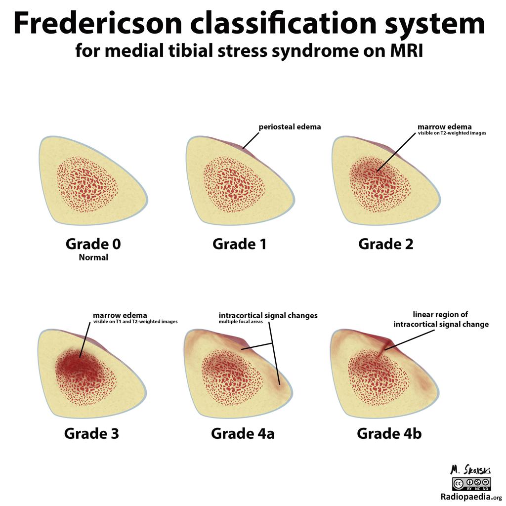
| 08:07, 13 February 2022 | Medial tibial stress Fredericson grading.jpg (file) |  |
115 KB | Jeremy | From https://radiopaedia.org/cases/21292/studies/21209?lang=gb&referrer=%2Farticles%2Fmri-grading-system-for-bone-stress-injuries%3Flang%3Dgb%23image_list_item_2851866 | 1 |
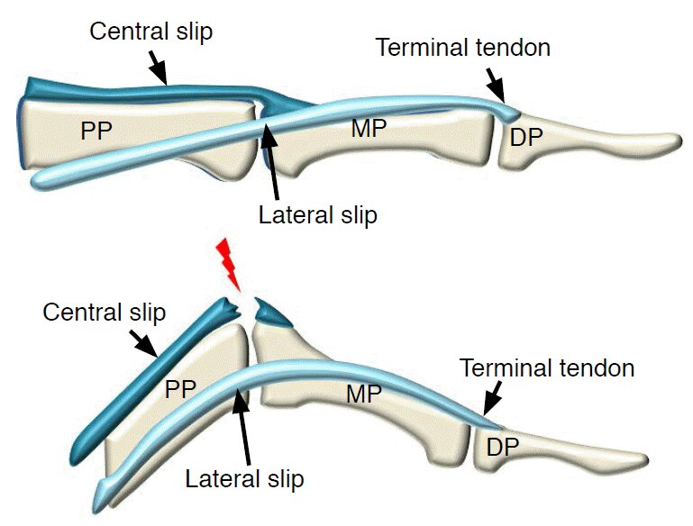
| 07:36, 7 February 2022 | Boutonniere deformity.jpg (file) |  |
58 KB | Jeremy | Schematic images of Boutonnière deformity. Boutonnière deformity is an injury involving the central slip of the extensor tendon over the proximal interphalangeal joint. DP, distal phalanx; MP, middle phalanx; PP, proximal phalanx. Lee SA, Kim BH, Kim SJ, Kim JN, Park SY, Choi K. Current status of ultrasonography of the finger. Ultrasonography. 2016 Apr;35(2):110-23. doi: 10.14366/usg.15051. Epub 2015 Nov 24. PMID:26753604; PMCID: PMC4825212. | 1 |
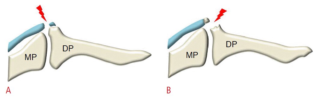
| 07:35, 7 February 2022 | Mallet finger.jpg (file) |  |
28 KB | Jeremy | A. Mallet finger is a tear of the extensor tendon at the insertion of the terminal tendon into the distal phalangeal base. B. The tendon may pull off a piece of bone, suggestive of an avulsion fracture. DP, distal phalanx; MP, middle phalanx. Lee SA, Kim BH, Kim SJ, Kim JN, Park SY, Choi K. Current status of ultrasonography of the finger. Ultrasonography. 2016 Apr;35(2):110-23. doi: 10.14366/usg.15051. Epub 2015 Nov 24. PMID:26753604; PMCID: PMC4825212. | 1 |
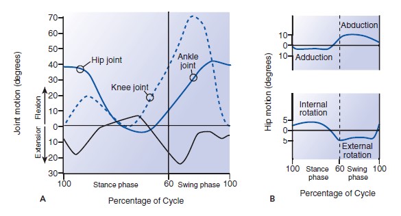
| 07:05, 7 February 2022 | Range of motion with gait.jpg (file) |  |
42 KB | Jeremy | Nordin, Margareta, and Victor H. Frankel. Basic biomechanics of the musculoskeletal system. Philadelphia: Wolters Kluwer Health/Lippincott Williams & Wilkins, 2012. | 1 |
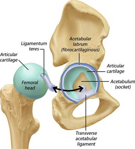
| 06:51, 7 February 2022 | Acetabular-labrum-picture.jpg (file) |  |
23 KB | Jeremy | Source unknown | 1 |
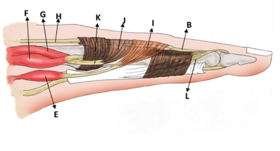
| 12:50, 6 February 2022 | Finger extensor mechanism lateral view.jpg (file) |  |
21 KB | Jeremy | a: triangular ligament; b: central slip; c: slips of long extensor tendon to lateral bands; d: lateral bands; e: lumbrical muscle; f: superficial belly of dorsal interosseous muscle; g: deep belly of dorsal interosseous muscle or palmar interosseous muscle; h: long extensor tendon; i: oblique fibers; j: transverse fibers; k: saggital bands; l: oblique retinacular ligament (Landmeer’s ligament) Ditsios K, Konstantinou P, Pinto I, Karavelis A, Kostretzis L (2017) Extensor Mechanism’s Anatomy a... | 1 |
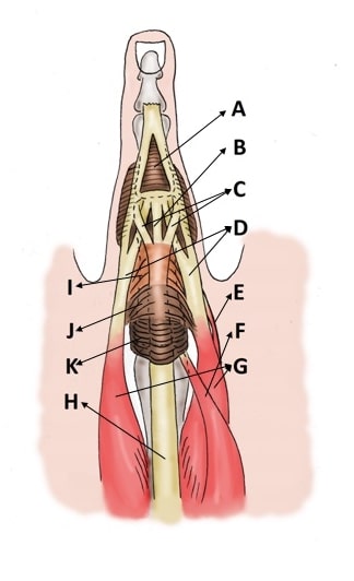
| 12:50, 6 February 2022 | Finger extensor mechanism dorsal view.jpg (file) |  |
23 KB | Jeremy | a: triangular ligament; b: central slip; c: slips of long extensor tendon to lateral bands; d: lateral bands; e: lumbrical muscle; f: superficial belly of dorsal interosseous muscle; g: deep belly of dorsal interosseous muscle or palmar interosseous muscle; h: long extensor tendon; i: oblique fibers; j: transverse fibers; k: saggital bands; l: oblique retinacular ligament (Landmeer’s ligament) Ditsios K, Konstantinou P, Pinto I, Karavelis A, Kostretzis L (2017) Extensor Mechanism’s Anatomy a... | 1 |
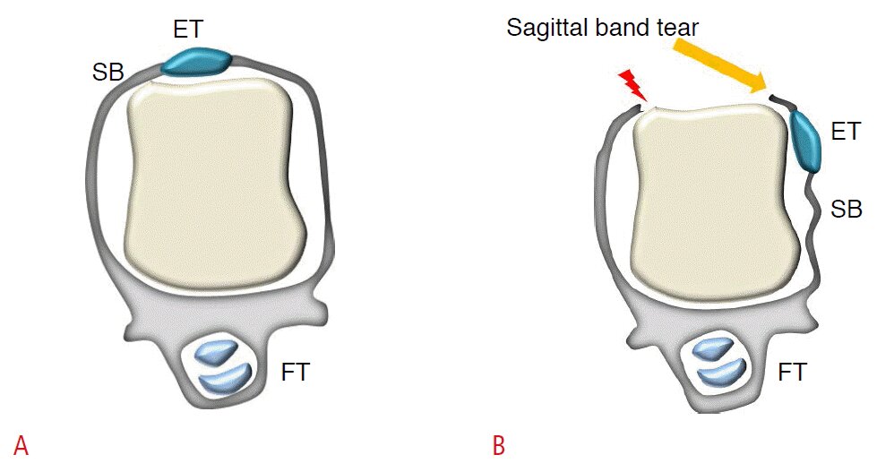
| 12:34, 6 February 2022 | Sagittal band tear extensor tendon subluxation.mp4 (file) | 1.21 MB | Jeremy | Sagittal band injury. Since the sagittal band prevents deviation of the extensor tendon during metacarpophalangeal joint flexion and bowstring during hyperextension, sagittal band injury causes extensor tendon dislocation during flexion.<ref>Lee SA, Kim BH, Kim SJ, Kim JN, Park SY, Choi K. Current status of ultrasonography of the finger. Ultrasonography. 2016 Apr;35(2):110-23. doi: 10.14366/usg.15051. Epub 2015 Nov 24. PMID:26753604; PMCID: PMC4825212.</ref> | 1 | |
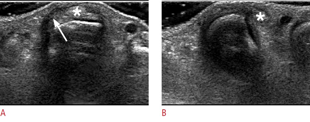
| 12:30, 6 February 2022 | Sagittal band tear ultrasound.jpg (file) |  |
82 KB | Jeremy | Sagittal band tear in a 43-year-old woman. A. A transverse sonogram of the third metacarpophalangeal joint when the finger is extended shows an abnormal radial sagittal band with irregularity and hypoechogenicity (arrow). The extensor tendon (asterisk) is positioned normally. B. A dynamic examination obtained in the transverse plane during finger flexion shows dislocation of the extensor tendon (asterisk).<ref>Lee SA, Kim BH, Kim SJ, Kim JN, Park SY, Choi K. Current status of ultrasonography... | 1 |
| 12:24, 6 February 2022 | Sagittal band tear.jpg (file) |  |
54 KB | Jeremy | Lee SA, Kim BH, Kim SJ, Kim JN, Park SY, Choi K. Current status of ultrasonography of the finger. Ultrasonography. 2016 Apr;35(2):110-23. doi: 10.14366/usg.15051. Epub 2015 Nov 24. PMID: 26753604; PMCID: PMC4825212. | 1 |
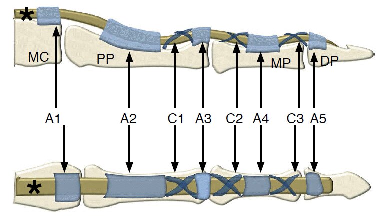
| 07:41, 6 February 2022 | Finger pulley system.jpg (file) |  |
47 KB | Jeremy | Lee SA, Kim BH, Kim SJ, Kim JN, Park SY, Choi K. Current status of ultrasonography of the finger. Ultrasonography. 2016 Apr;35(2):110-23. doi: 10.14366/usg.15051. Epub 2015 Nov 24. PMID: 26753604; PMCID: PMC4825212. | 1 |
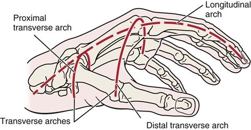
| 07:28, 6 February 2022 | Hand arches.jpg (file) |  |
32 KB | Jeremy | Unknown source | 1 |
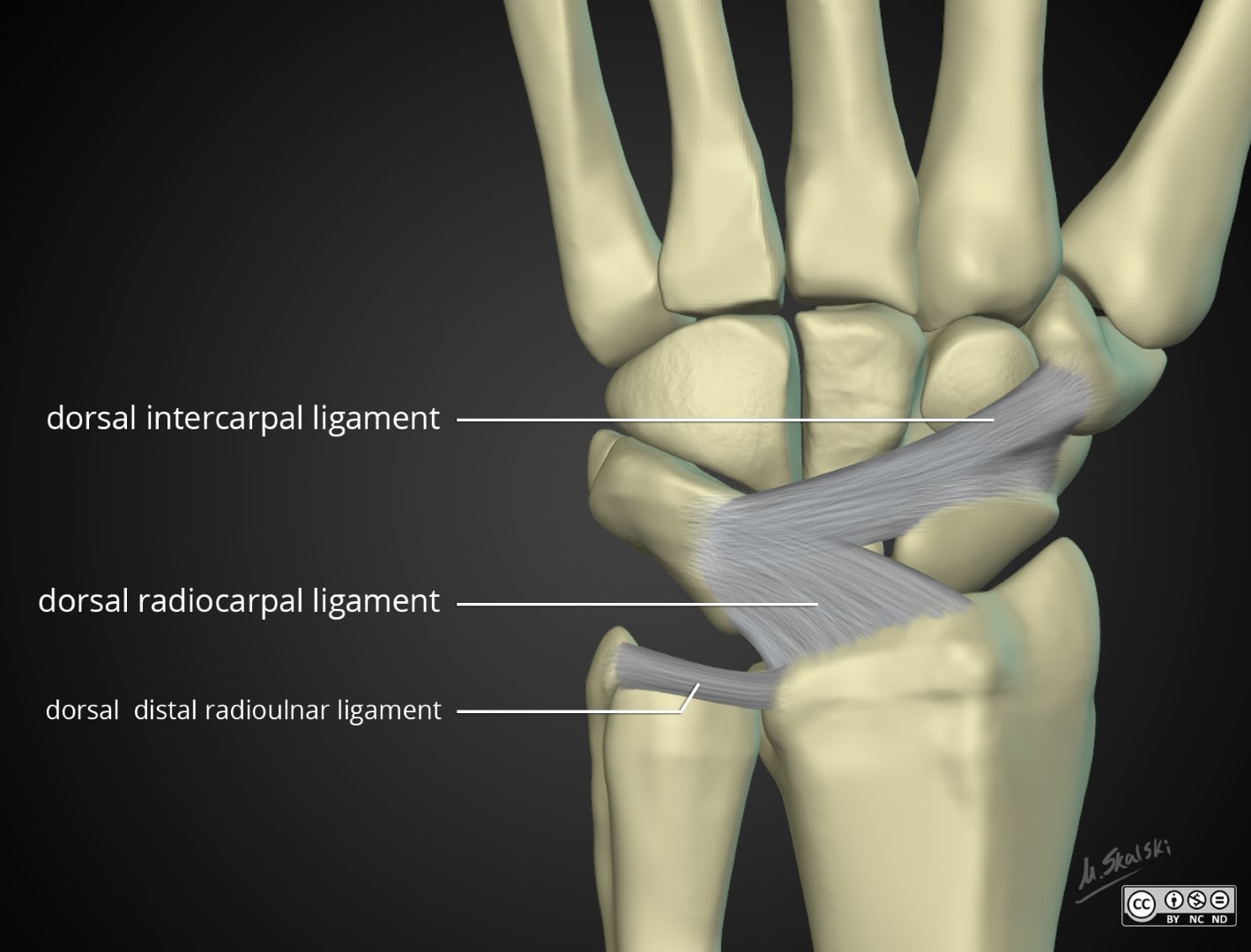
| 07:22, 6 February 2022 | Wrist-anatomy-extrinsic-ligaments dorsal.jpg (file) |  |
90 KB | Jeremy | Case courtesy of Dr Matt Skalski, <a href="https://radiopaedia.org/?lang=gb">Radiopaedia.org</a>. From the case <a href="https://radiopaedia.org/cases/43845?lang=gb">rID: 43845</a> | 1 |
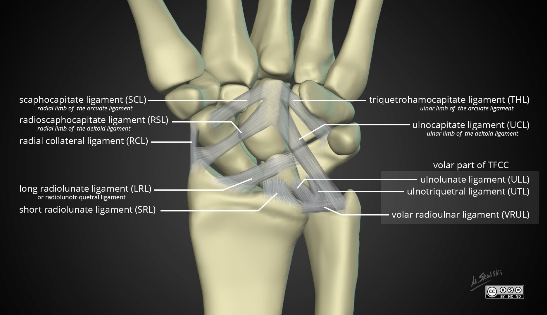
| 07:18, 6 February 2022 | Wrist-anatomy-extrinsic-ligaments.jpg (file) |  |
150 KB | Jeremy | Case courtesy of Dr Matt Skalski, <a href="https://radiopaedia.org/?lang=gb">Radiopaedia.org</a>. From the case <a href="https://radiopaedia.org/cases/43845?lang=gb">rID: 43845</a> | 1 |
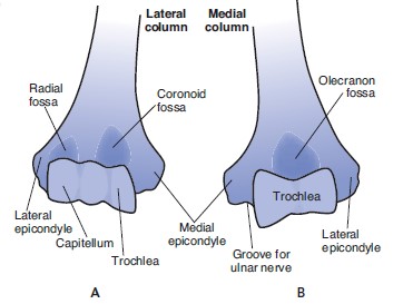
| 06:49, 6 February 2022 | Distal humerus medial and lateral columns.jpg (file) |  |
23 KB | Jeremy | Nordin, Margareta, and Victor H. Frankel. Basic biomechanics of the musculoskeletal system. Philadelphia: Wolters Kluwer Health/Lippincott Williams & Wilkins, 2012. | 1 |
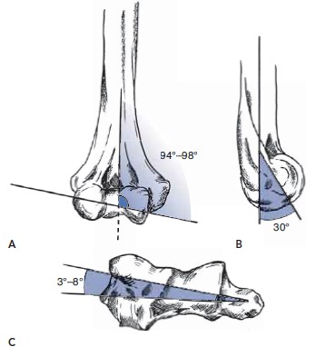
| 06:46, 6 February 2022 | Distal humerus angulations.jpg (file) |  |
25 KB | Jeremy | Copyright Nordin and Frankel Basic BIomechanics of the Musculoskeletal System 4th edition | 1 |
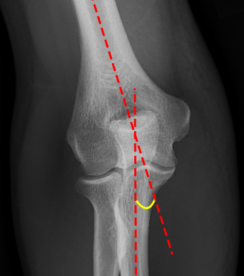
| 06:38, 6 February 2022 | Normal-elbow-carrying-angle.jpg (file) |  |
123 KB | Jeremy | Case courtesy of Dr Samir Benoudina, <a href="https://radiopaedia.org/?lang=gb">Radiopaedia.org</a>. From the case <a href="https://radiopaedia.org/cases/42315?lang=gb">rID: 42315</a> | 1 |
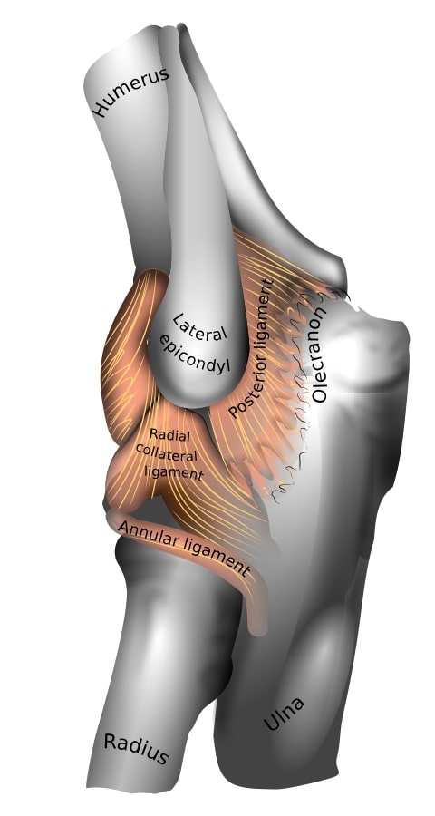
| 06:19, 6 February 2022 | Elbow joint lateral ligaments.jpg (file) |  |
43 KB | Jeremy | https://en.wikipedia.org/wiki/File:En-elbow_joint.svg | 1 |
| 20:35, 31 January 2022 | NZCMM Training Manual Dec 2021.pdf (file) | 429 KB | Jeremy | 1 | ||
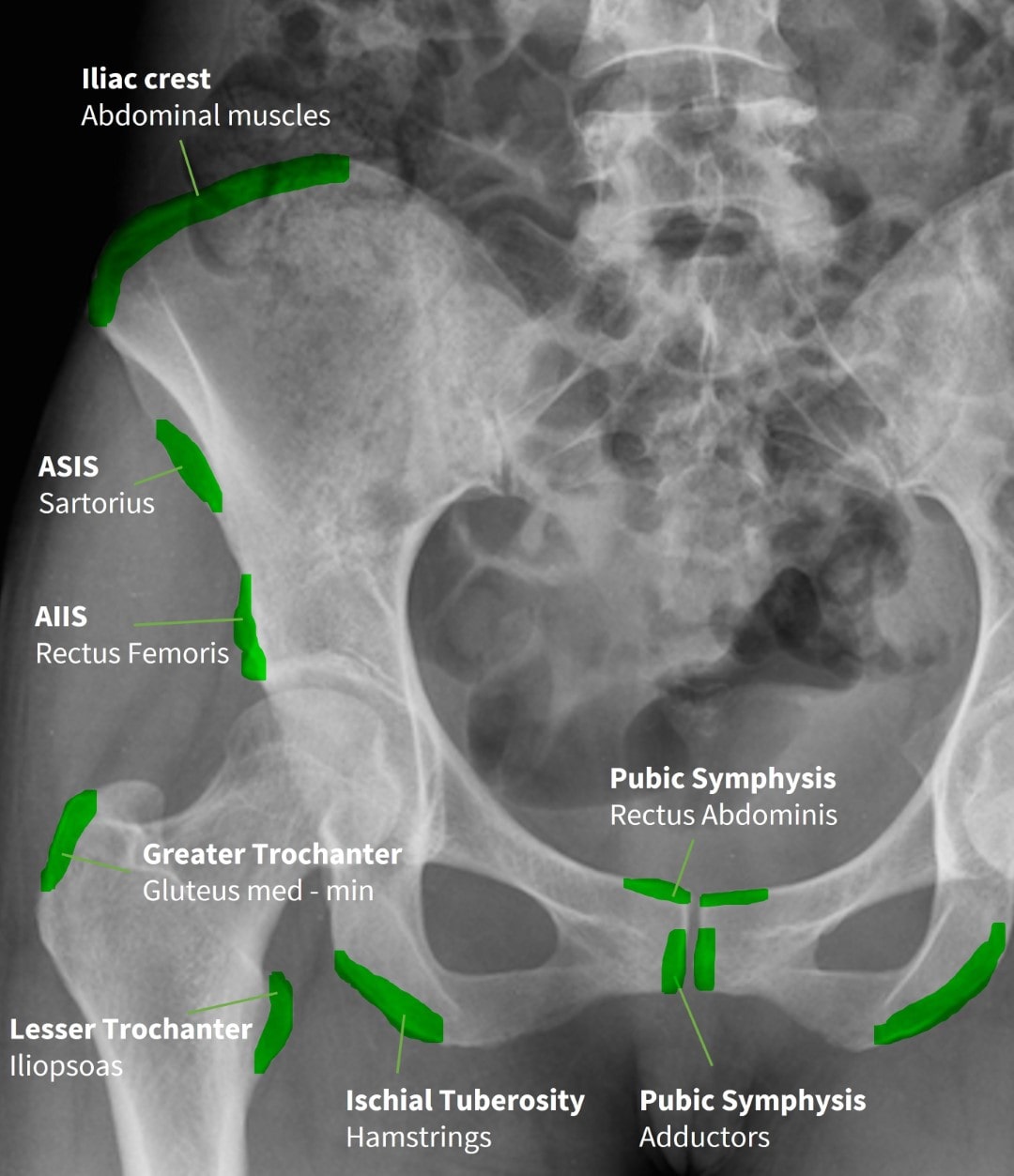
| 15:53, 16 January 2022 | Pelvis and Hip Radiograph Muscle Attachments .jpg (file) |  |
136 KB | Jeremy | 1 | |
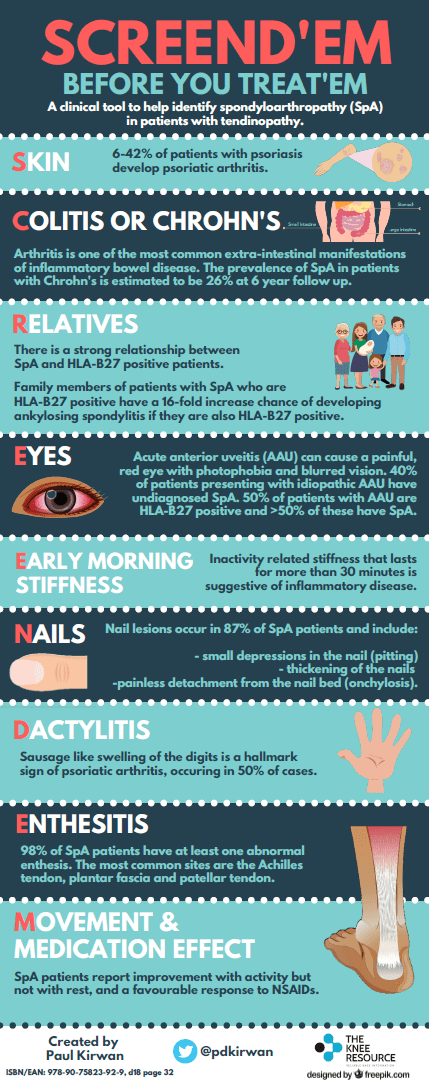
| 15:12, 16 January 2022 | Screendem.png (file) |  |
71 KB | Jeremy | From @pdkirwan | 1 |
| 09:16, 16 January 2022 | Jeremy S.jpg (file) |  |
129 KB | Jeremy | 1 | |
| 17:55, 12 January 2022 | Needle bend.jpg (file) | 35 KB | Jeremy | 1 | ||
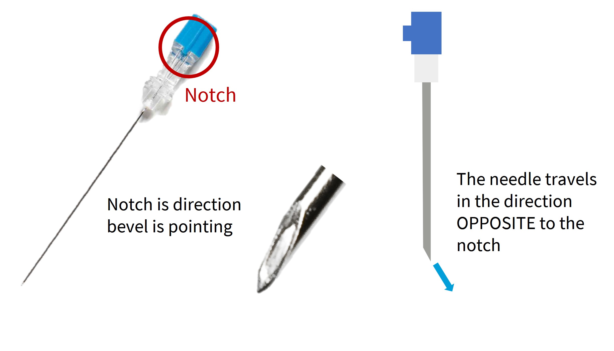
| 17:30, 12 January 2022 | Needle bevel direction.jpg (file) |  |
169 KB | Jeremy | 1 | |
| 20:05, 6 January 2022 | NZCMM-logo-long.png (file) | 26 KB | Jeremy | 2 | ||
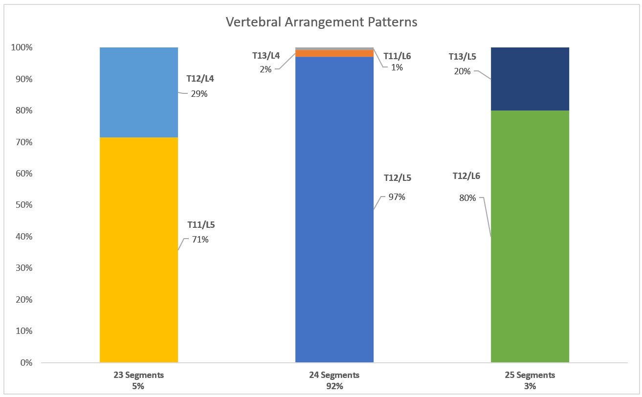
| 19:56, 6 January 2022 | Vertebral Arrangement Patterns.jpg (file) |  |
69 KB | Jeremy | 2 | |
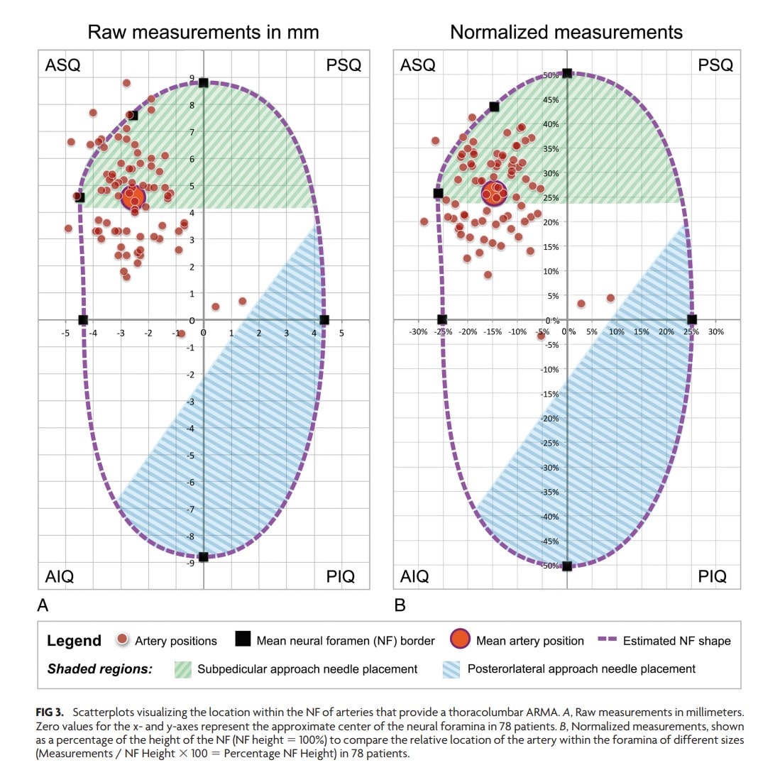
| 18:27, 4 January 2022 | Scatterplot of arteries in the neural foramen.jpg (file) |  |
207 KB | Jeremy | Copyrighted from open access article Gregg L, Sorte DE, Gailloud P. Intraforaminal Location of Thoracolumbar Radicular Arteries Providing an Anterior Radiculomedullary Artery Using Flat Panel Catheter Angiotomography. AJNR Am J Neuroradiol. 2017 May;38(5):1054-1060. doi: 10.3174/ajnr.A5104. Epub 2017 Feb 16. PMID: 28209578; PMCID: PMC7960376. | 1 |
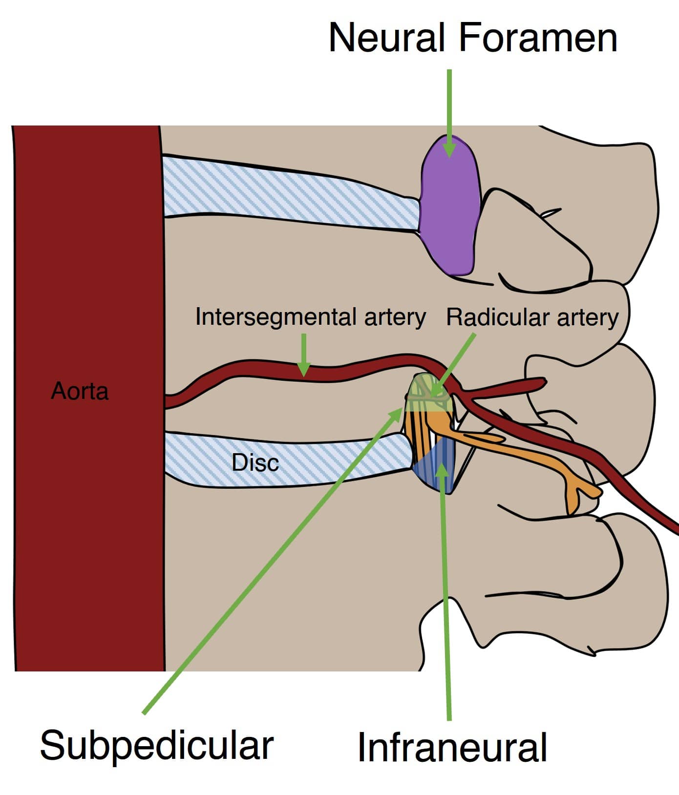
| 18:19, 4 January 2022 | Oblique view lumbar foramen.jpg (file) |  |
99 KB | Jeremy | 1 | |
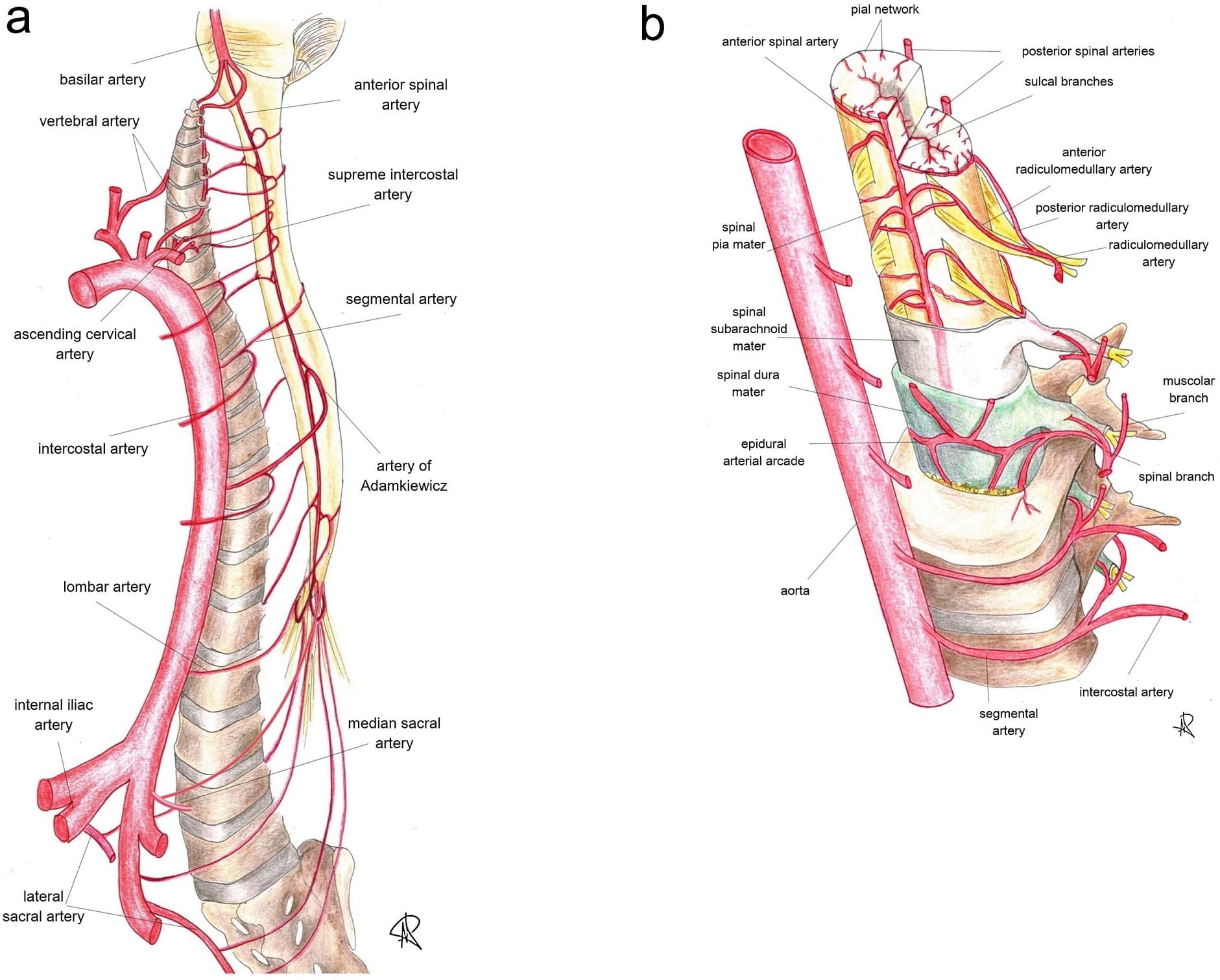
| 17:33, 4 January 2022 | Spinal cord blood supply.jpg (file) |  |
241 KB | Jeremy | Fig. 2. (a and b). Sectional representation of the arterial blood supply and feeling vessels to spinal cord (see the text for further details), whose catheterization is mandatory during spinal angiography. Along the cranio-caudal direction, the angiographic study should include and demonstrate: vertebral arteries, ascending cervical arteries, deep cervical arteries (not shown), supreme intercostal arteries, intercostal arteries, lumbar arteries, median and lateral sacral arteries. Identifica... | 1 |
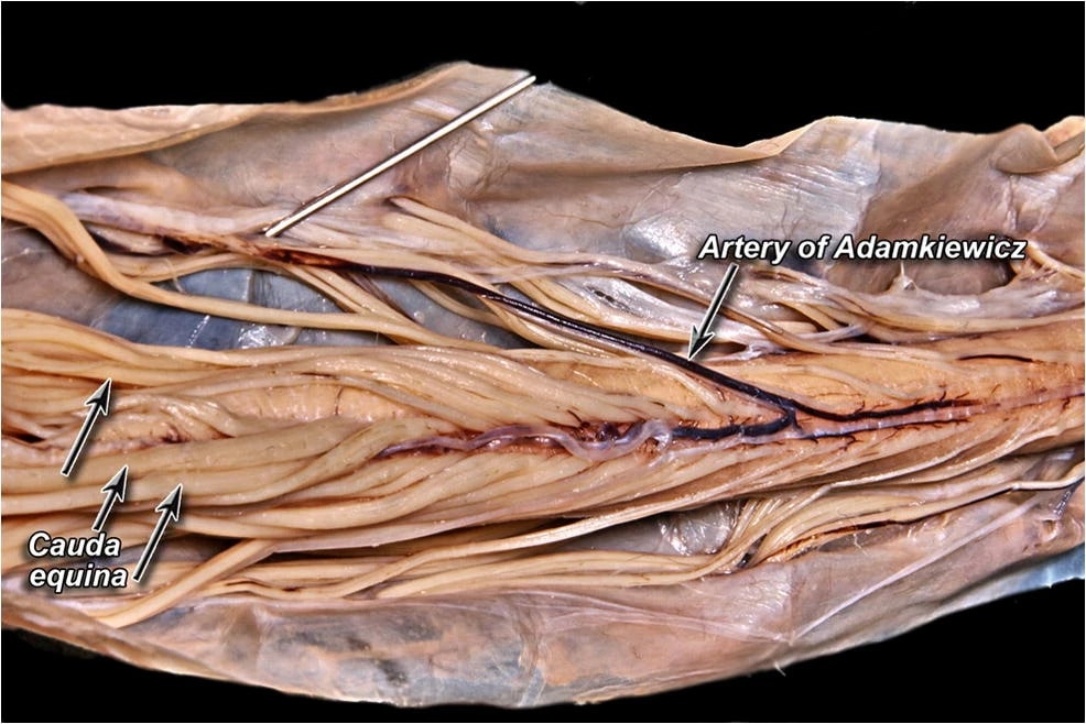
| 17:16, 4 January 2022 | Cadaveric-dissection-of-the-artery-of-Adamkiewicz.jpeg (file) |  |
128 KB | Jeremy | From Taterra D, Skinningsrud B, Pękala PA, Hsieh WC, Cirocchi R, Walocha JA, Tubbs RS, Tomaszewski KA, Henry BM. Artery of Adamkiewicz: a meta-analysis of anatomical characteristics. Neuroradiology. 2019 Aug;61(8):869-880. doi: 10.1007/s00234-019-02207-y. Epub 2019 Apr 27. PMID: 31030251; PMCID: PMC6620248. | 1 |
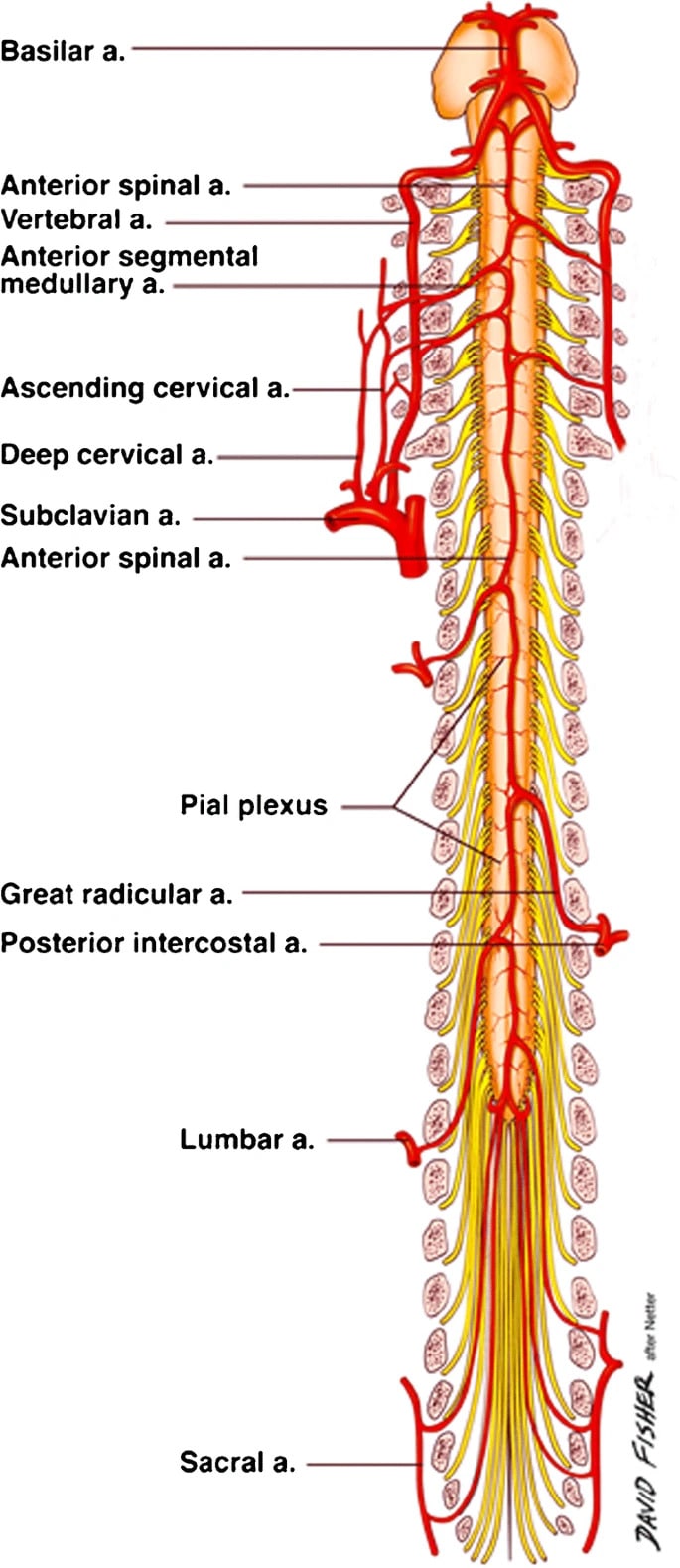
| 16:17, 4 January 2022 | Spinal Cord Vasculature.jpeg (file) |  |
134 KB | Jeremy | From Taterra D, Skinningsrud B, Pękala PA, Hsieh WC, Cirocchi R, Walocha JA, Tubbs RS, Tomaszewski KA, Henry BM. Artery of Adamkiewicz: a meta-analysis of anatomical characteristics. Neuroradiology. 2019 Aug;61(8):869-880. doi: 10.1007/s00234-019-02207-y. Epub 2019 Apr 27. PMID: 31030251; PMCID: PMC6620248. | 1 |
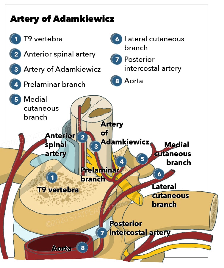
| 15:51, 4 January 2022 | Artery of Adamkiewicz.jpg (file) |  |
121 KB | Jeremy | From https://www.ncbi.nlm.nih.gov/books/NBK532971/ | 1 |
| 15:45, 4 January 2022 | Coronal spinal nerve relationship.jpg (file) |  |
132 KB | Jeremy | 3 | |
| 06:09, 4 January 2022 | Anatomy and Clinical Importance of the Epidural Space - Fyneface-Ogan 2012.pdf (file) | 521 KB | Jeremy | 1 | ||
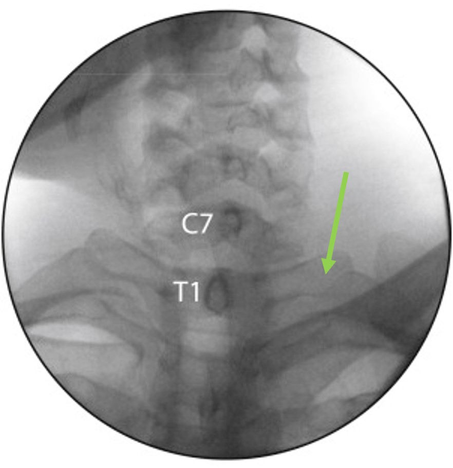
| 13:35, 3 January 2022 | Cervicothoracic junction AP fluoroscopy.jpg (file) |  |
70 KB | Jeremy | The transverse processes of T1 are upsloping and longer than the transverse processes of C7 | 1 |
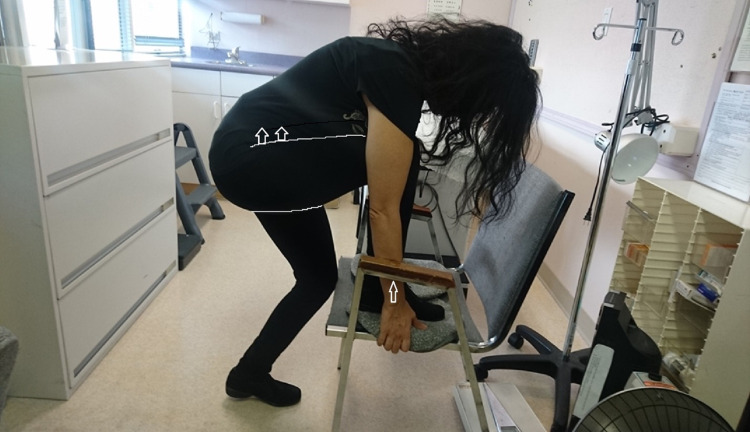
| 12:26, 18 December 2021 | SIFFT-E to level a right anterior sacroiliac malrotation.jpg (file) |  |
87 KB | Jeremy | SIFFT-E treating a right anterior sacroiliac torsion: The right thigh pushes the right ASIS backward Place the right foot on the seat of the chair with the hands holding the seat on either side of the foot. The left knee touches the front of the seat. Lean back. Pull up hard with both hands on the seat of the chair and hold for a full two minutes. Six of the 62 participants did the chair exercise. It indicated when the only tender PSIS is anteriorly displaced (higher than the other PSIS) and... | 1 |
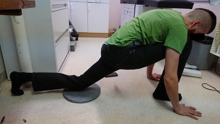
| 12:25, 18 December 2021 | SIFFT-E to level a right anterior left posterior sacroiliac malrotation.jpg (file) |  |
83 KB | Jeremy | SIFFT-E stretch exercise to level a right anterior, left posterior sacroiliac malrotation Place the right foot and the left knee on the floor hands on either side of the right foot. Lean forward so that the right thigh is pushing up hard on the right ASIS to level an anterior SI torsion. Slide the left knee as far back as possible to hyperextend the left thigh. A strong pull should be felt in the left groin to level a posterior SI torsion. Hold this position for a full two minutes. If only on... | 1 |
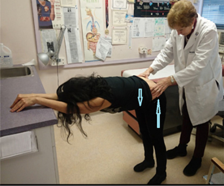
| 12:10, 18 December 2021 | Sacroiliac Forward Flexion Test.jpg (file) |  |
104 KB | Jeremy | Sacroiliac forward flexion test (SIFFT) The patient is leaning against the desk to relax the buttock muscles, making them softer and easier to press down on. The legs push the PSISs up, while the weight of the body brings the spine down, which rotates the sacrum anteriorly making the PSISs easier to feel. PSISs are located by pressing down firmly with the ulnar side of the thumbs while gradually advancing the cephalad toward the PSISs starting on either side of the intergluteal cleft. When th... | 1 |
| 11:06, 14 December 2021 | Fibromyalgia Diagnostic Criteria 2010.pdf (file) | 219 KB | Jeremy | 1 | ||
| 05:20, 10 December 2021 | Budapest-Criteria-Ver-3.0.pdf (file) | 443 KB | Jeremy | 1 | ||
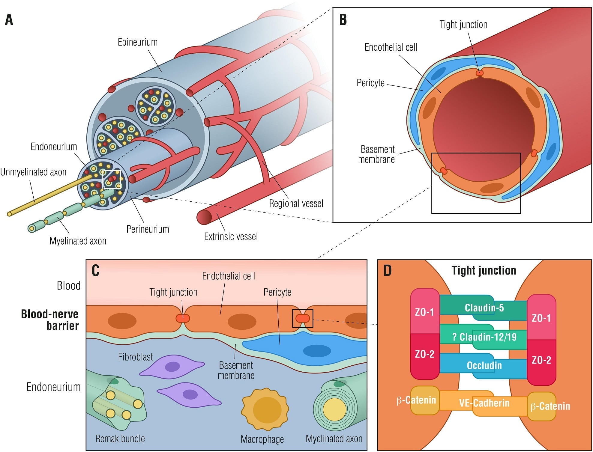
| 03:08, 6 December 2021 | Blood nerve barrier.jpg (file) |  |
263 KB | Jeremy | Figure 1. Blood-nerve barrier. (A) Transverse view of a peripheral nerve ensheathed by epineurial collagen fibrils (epineurium) and blood vessels. Individual nerve fascicles consisting of unmyelinated and myelinated axons as well as small blood vessels are ensheathed by the perineurium, forming the endoneurial microenvironment. (B) Individual endoneurial blood vessel surrounded by endothelial cells, pericytes and the basement membrane. (C) Cellular structure of the blood-nerve barrier, formed... | 1 |
| 03:03, 6 December 2021 | Carpal tunnel syndrome pathophysiology.png (file) |  |
151 KB | Jeremy | 2 | |
| 02:20, 6 December 2021 | Median nerve position variations wrist.png (file) |  |
96 KB | Jeremy | Source unknown | 1 |
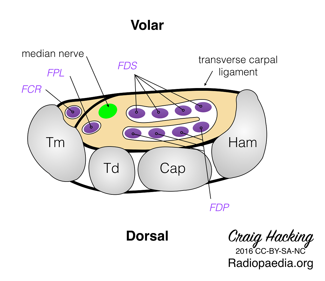
| 19:01, 5 December 2021 | Carpal-tunnel-diagram.png (file) |  |
93 KB | Jeremy | Case courtesy of Assoc Prof Craig Hacking, <a href="https://radiopaedia.org/?lang=gb">Radiopaedia.org</a>. From the case <a href="https://radiopaedia.org/cases/47155?lang=gb">rID: 47155</a> | 1 |
| 08:17, 30 October 2021 | NZCMM Training Manual Oct 2021.pdf (file) | 514 KB | Jeremy | File uploaded with MsUpload | 1 |

