File list
From WikiMSK
This special page shows all uploaded files.
| Date | Name | Thumbnail | Size | User | Description | Versions |
|---|---|---|---|---|---|---|
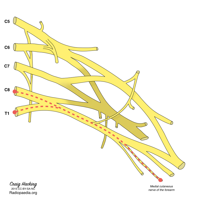
| 08:53, 23 April 2022 | Medial antebrachial cutaneous nerve.png (file) |  |
154 KB | Jeremy | From https://radiopaedia.org/articles/medial-cutaneous-nerve-of-the-forearm | 1 |
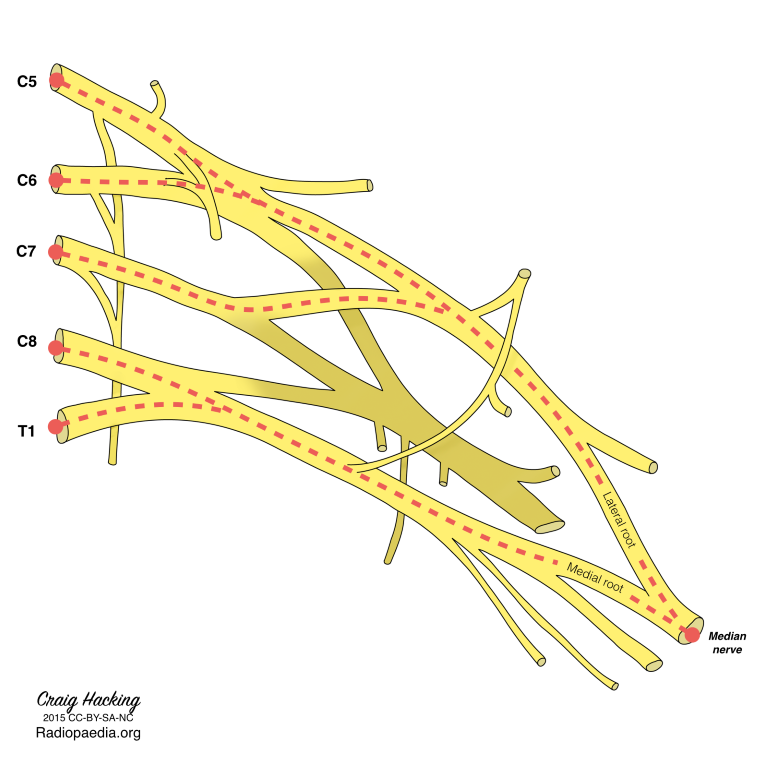
| 08:49, 23 April 2022 | Median nerve.png (file) |  |
168 KB | Jeremy | From https://radiopaedia.org/articles/median-nerve-2?lang=gb | 1 |
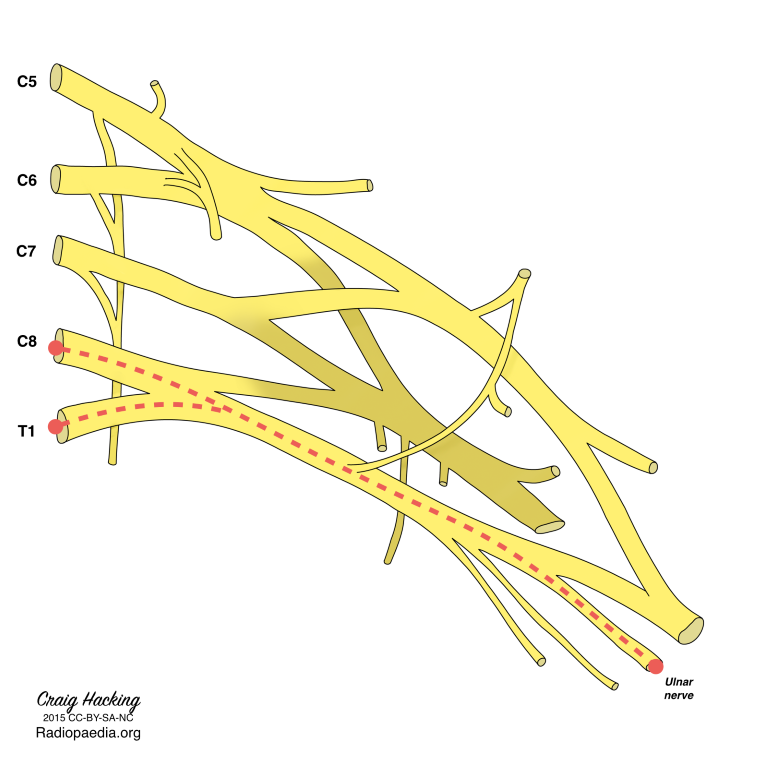
| 08:47, 23 April 2022 | Ulnar nerve.png (file) |  |
154 KB | Jeremy | From https://radiopaedia.org/articles/ulnar-nerve | 1 |
| 08:08, 23 April 2022 | Myofascial Chains Systematic Review - Wilke 2016.pdf (file) | 574 KB | Jeremy | File uploaded with MsUpload | 1 | |
| 07:54, 23 April 2022 | Quadrilateral Space Syndrome - Hangge 2018.pdf (file) | 1.2 MB | Jeremy | 1 | ||
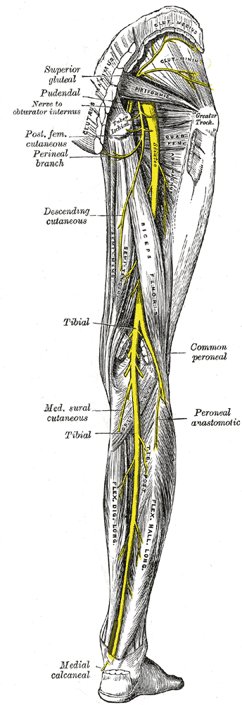
| 08:23, 18 April 2022 | Posterior leg nerves Gray832.png (file) |  |
70 KB | Jeremy | 1 | |
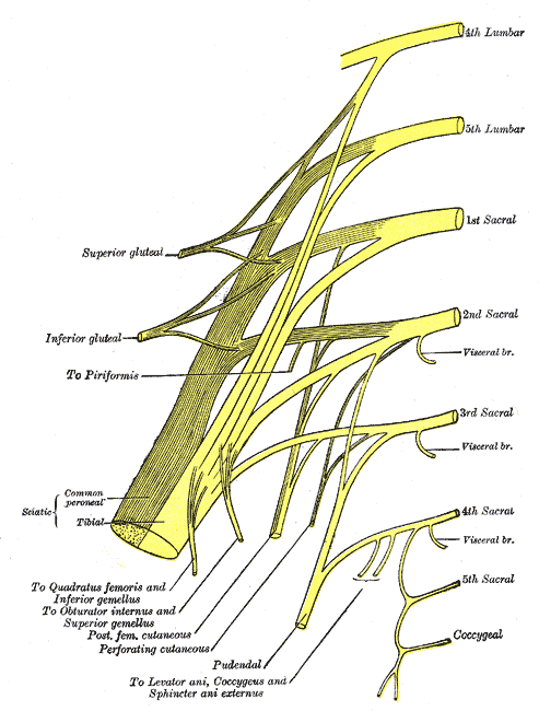
| 08:22, 18 April 2022 | Sacral and pudendal plexuses Gray828.png (file) |  |
34 KB | Jeremy | 1 | |
| 22:23, 17 April 2022 | Mucular cramps cause and management - Swash 2019.pdf (file) | 246 KB | Jeremy | Uploaded with SimpleBatchUpload | 1 | |
| 22:14, 17 April 2022 | Cartilage in normal and osteoarthritis conditions - Martel-Pelletier 2008.pdf (file) | 356 KB | Jeremy | Uploaded with SimpleBatchUpload | 1 | |
| 22:09, 17 April 2022 | MRI of the knee - Hash 2013.pdf (file) | 4.63 MB | Jeremy | Uploaded with SimpleBatchUpload | 1 | |
| 22:06, 17 April 2022 | Hip and Knee Strengthening in Patellofemoral Syndrome Metaanalysis - Nascimiento 2018.pdf (file) | 477 KB | Jeremy | File uploaded with MsUpload | 1 | |
| 22:01, 17 April 2022 | Femoroacetabular Impingement Radiographic Diagnosis - Tannast 2006.pdf (file) | 3.12 MB | Jeremy | Uploaded with SimpleBatchUpload | 1 | |
| 22:00, 17 April 2022 | Imaging of Femoral Acetabular impingement sydrome - Manaster 2006.pdf (file) | 794 KB | Jeremy | Uploaded with SimpleBatchUpload | 1 | |
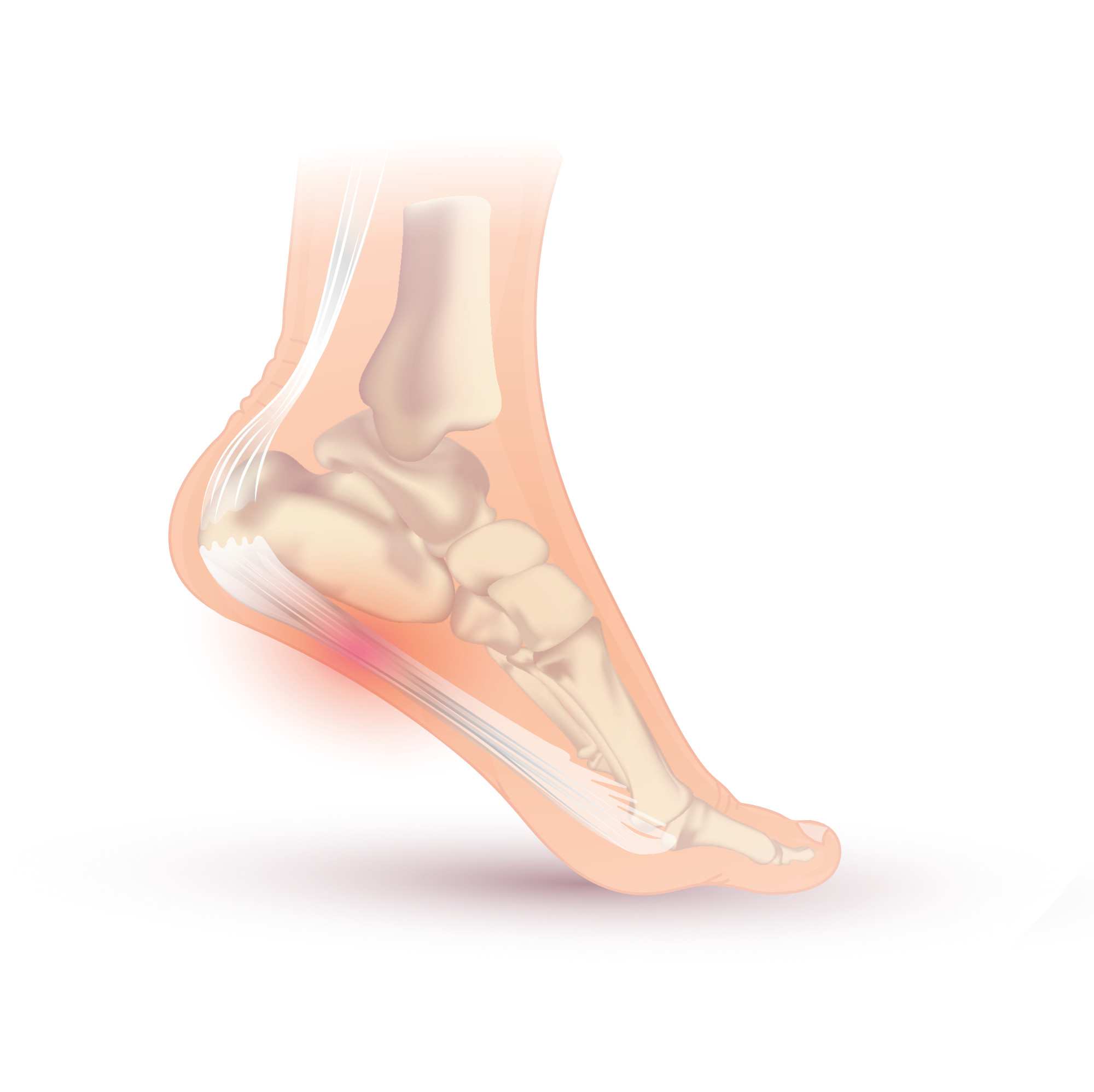
| 21:57, 17 April 2022 | Achilles tendon disorders - Asplund 2013.pdf (file) | 616 KB | Jeremy | Uploaded with SimpleBatchUpload | 1 | |
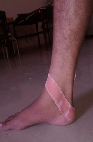
| 21:54, 17 April 2022 | Calcaneal taping.jpg (file) |  |
20 KB | Jeremy | From https://www.ssjournals.com/index.php/ijbar/article/view/2303 | 1 |
| 21:37, 17 April 2022 | Physiotherapy management of patellar tendinopathy J Cook.pdf (file) | 1.36 MB | Jeremy | File uploaded with MsUpload | 1 | |
| 19:23, 16 April 2022 | Plantar flexion inversion and dorsiflexion and eversion tarsal tunnel.jpg (file) |  |
32 KB | Jeremy | Reproduced with permission from A.M. Trescot (ed.), Peripheral Nerve Entrapments: Clinical Diagnosis and Management, DOI 10.1007/978-3-319-27482-9_74 | 1 |
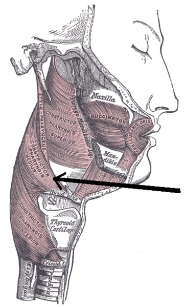
| 17:02, 16 April 2022 | Musculusconstrictorpharyngismedius.jpg (file) |  |
46 KB | Jeremy | 1 | |
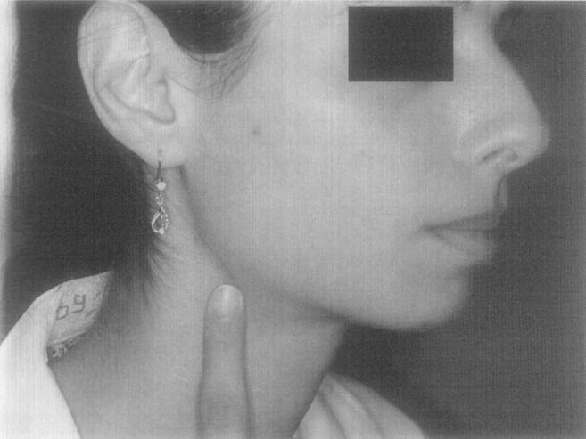
| 16:25, 16 April 2022 | Hyoid syndrome tenderness.jpg (file) |  |
66 KB | Jeremy | From Nir D, Hefer T, Joachims HZ. Hyoid bone syndrome and its treatment with nonsteroidal anti-inflammatory drugs. Am J Otolaryngol. 1998 Sep-Oct;19(5):296-300. doi: 10.1016/s0196-0709(98)90001-1. PMID: 9758176. | 1 |
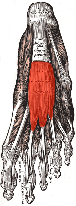
| 11:36, 16 April 2022 | Flexor digitorum brevis.png (file) |  |
79 KB | Jeremy | 1 | |
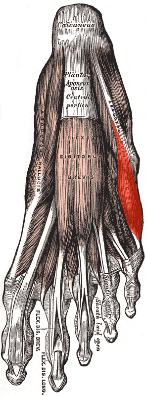
| 11:14, 16 April 2022 | Abductor digiti minimi (foot).png (file) | .png) |
75 KB | Jeremy | 1 | |
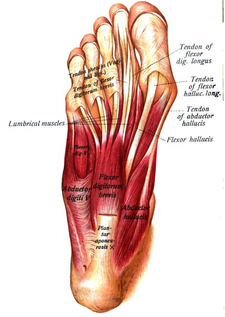
| 11:12, 16 April 2022 | Muscles plantar foot first layer Sobo.jpg (file) |  |
110 KB | Jeremy | 1 | |
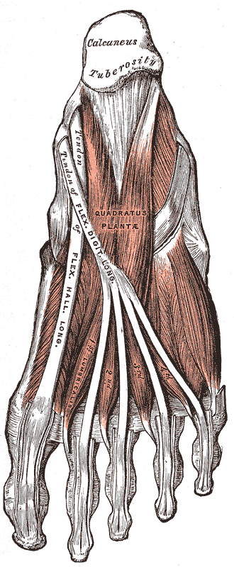
| 11:10, 16 April 2022 | Muscles plantar foot Gray444.png (file) |  |
59 KB | Jeremy | 1 | |
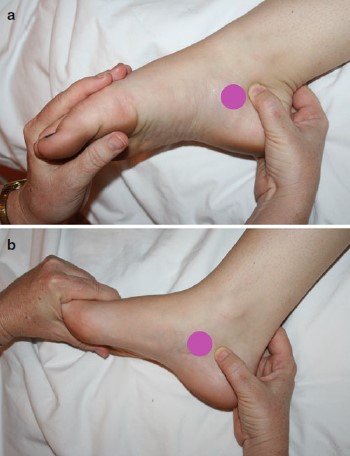
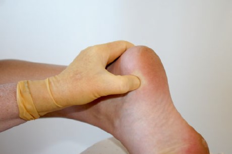
| 11:01, 16 April 2022 | Medial calcaneal tubercle tenderness.jpg (file) |  |
17 KB | Jeremy | With permission | 1 |
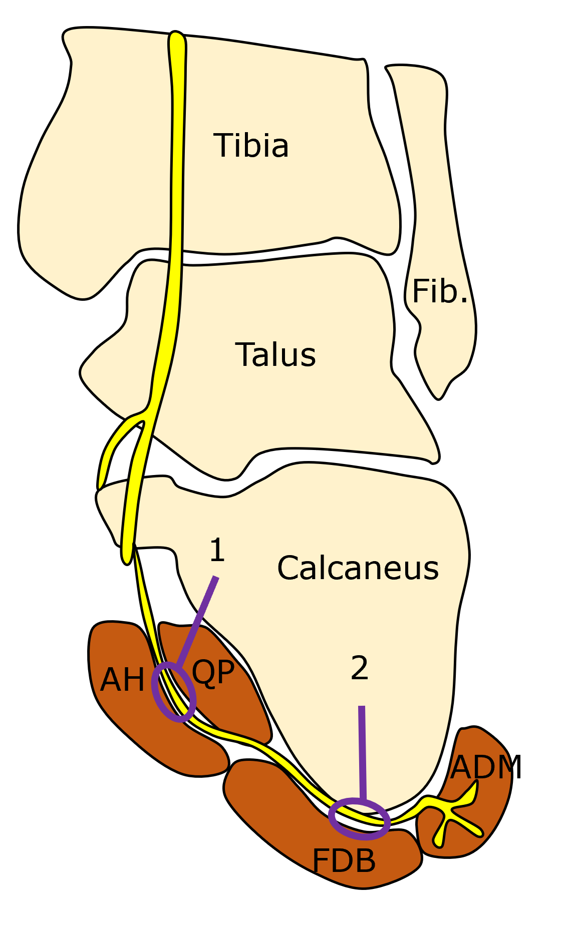
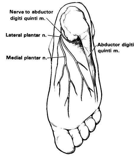
| 10:29, 16 April 2022 | Baxter nerve entrapment sites.png (file) |  |
200 KB | Jeremy | 1 | |
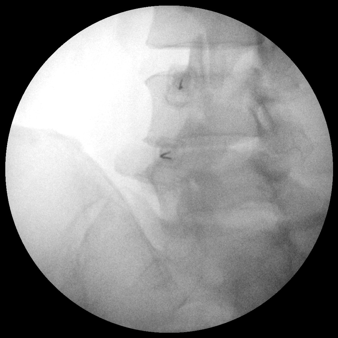
| 10:01, 16 April 2022 | Baxter nerve injection.mp4 (file) | 4.15 MB | Jeremy | From https://twitter.com/Dr_Ramon_Balius/status/711913973164146690?ref_src=twsrc%5Etfw%7Ctwcamp%5Etweetembed%7Ctwterm%5E711913973164146690%7Ctwgr%5E%7Ctwcon%5Es1_&ref_url=https%3A%2F%2Fwikimsk.org%2Fwiki%2FBaxter27s_Nerve_Entrapment | 1 | |
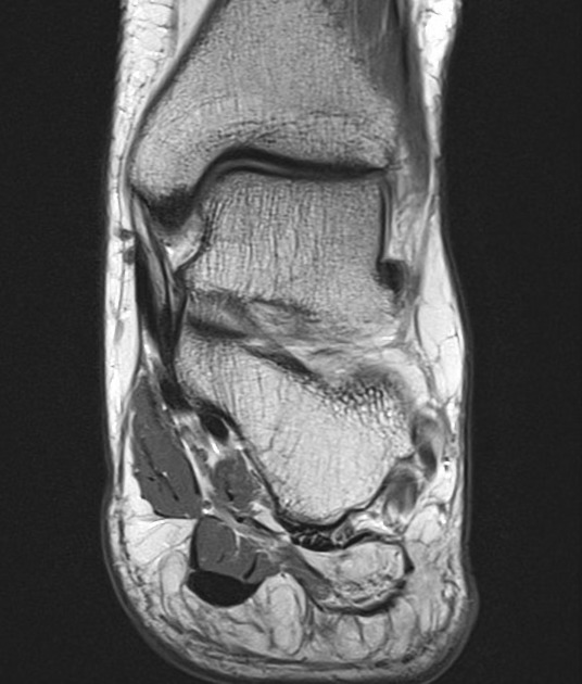
| 09:59, 16 April 2022 | Baxter nerve entrapment MRI.jpeg (file) |  |
67 KB | Jeremy | From Bauones, S., Feger, J. Baxter neuropathy. Reference article, Radiopaedia.org. (accessed on 15 Apr 2022) https://doi.org/10.53347/rID-25994 | 1 |
| 09:43, 16 April 2022 | Baxter nerve.jpg (file) |  |
44 KB | Jeremy | From Baxter DE, Thigpen CM. Heel pain--operative results. Foot Ankle. 1984 Jul-Aug;5(1):16-25. doi: 10.1177/107110078400500103. PMID: 6479759. | 1 |
| 21:53, 15 April 2022 | Plantar fasciitis.jpg (file) |  |
156 KB | Jeremy | From https://upload.wikimedia.org/wikipedia/commons/1/1a/Arch_tendonitis.jpg | 1 |
| 21:52, 15 April 2022 | Evaluation and Treatment of Chronic Plantar Fasciitis - Latt 2020.pdf (file) | 819 KB | Jeremy | File uploaded with MsUpload | 1 | |
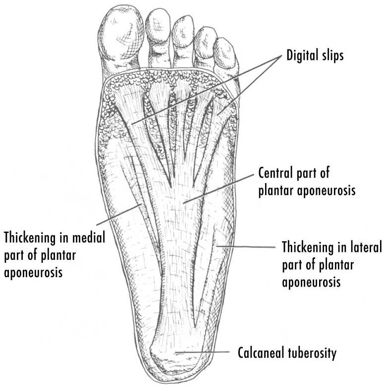
| 21:50, 15 April 2022 | Plantar fascia.jpg (file) |  |
69 KB | Jeremy | From https://upload.wikimedia.org/wikipedia/commons/b/b1/PF-PlantarDesignCrop.jpg | 1 |
| 20:48, 15 April 2022 | Shoe lifts for leg length discrepancy systematic review - Campbell 2017.pdf (file) | 355 KB | Jeremy | File uploaded with MsUpload | 1 | |
| 20:45, 15 April 2022 | Aetiology and Pathomechanics of FAI - Grantham 2019.pdf (file) | 837 KB | Jeremy | File uploaded with MsUpload | 1 | |
| 20:24, 15 April 2022 | Bone marrow oedema syndromes - Patel 2014.pdf (file) | 257 KB | Jeremy | File uploaded with MsUpload | 1 | |
| 20:03, 15 April 2022 | Femoral acetabular impingement FAI.svg (file) |  |
17 KB | Jeremy | From https://commons.wikimedia.org/wiki/File:Femoral_acetabular_impingement_FAI_de.svg | 1 |
| 13:57, 15 April 2022 | MBB Left L3 and L4 Oblique.jpg (file) |  |
141 KB | Jeremy | File uploaded with MsUpload | 1 |
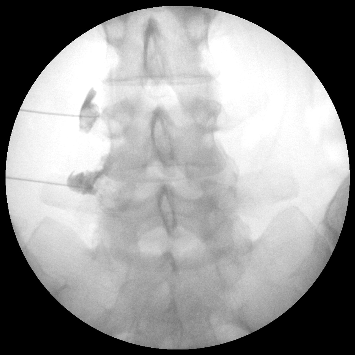
| 13:57, 15 April 2022 | MBB Left L3 and L4 AP contrast.jpg (file) |  |
115 KB | Jeremy | File uploaded with MsUpload | 1 |
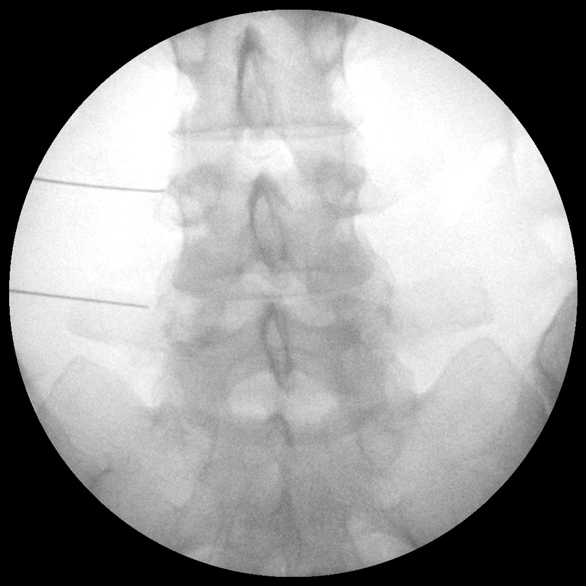
| 13:57, 15 April 2022 | MBB Left L3 and L4.jpg (file) |  |
139 KB | Jeremy | File uploaded with MsUpload | 1 |
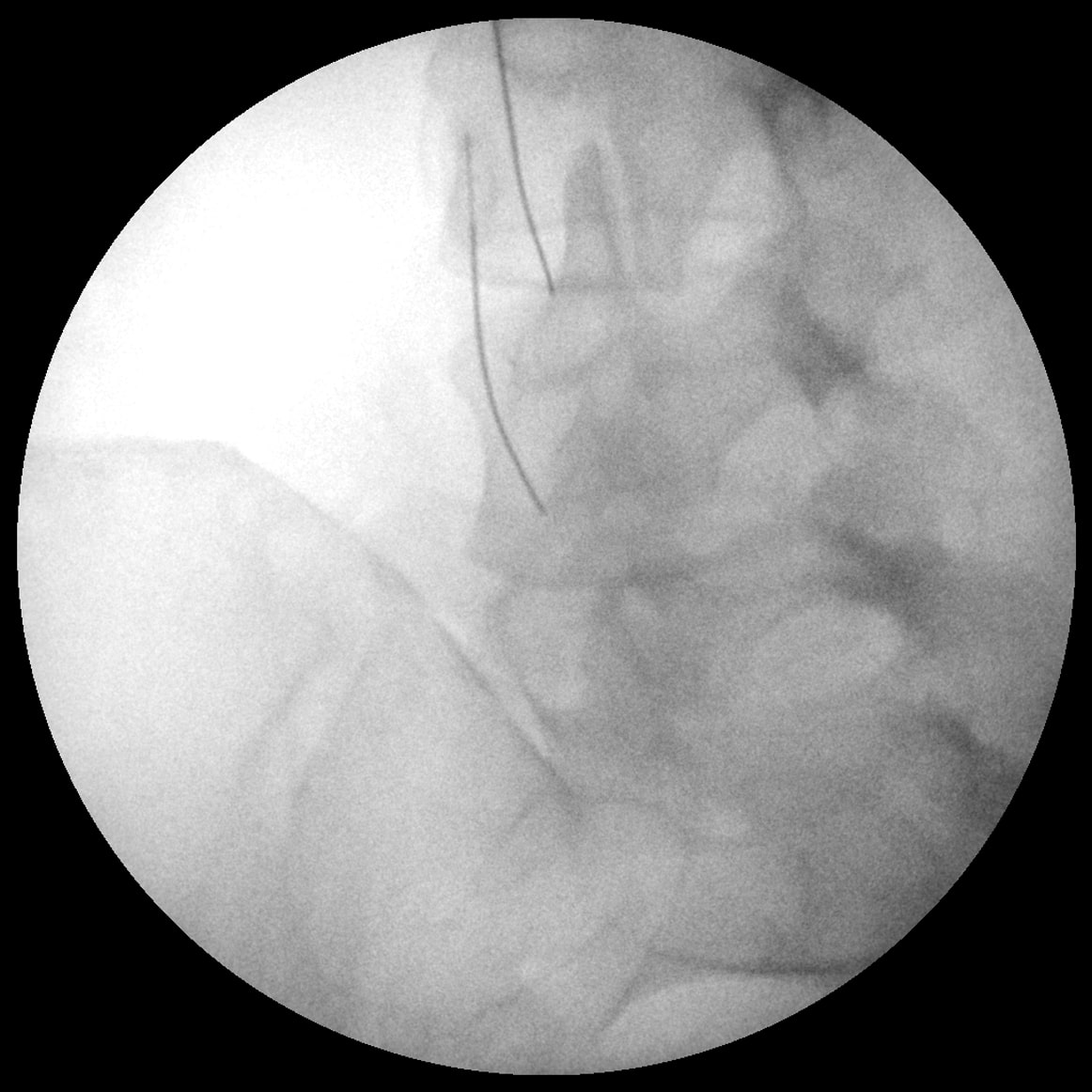
| 13:57, 15 April 2022 | MBB Left L3 and L4 Decline.jpg (file) |  |
144 KB | Jeremy | File uploaded with MsUpload | 1 |
| 08:51, 15 April 2022 | SIJ fluoroscopy oblique technique.jpg (file) |  |
41 KB | Jeremy | Oblique technique fluoroscopic view (A) and graphical illustration (B). In the oblique approach, the C-arm is rotated in a contralateral manner until the 2 joint lines become superimposed. Then one would target the inferior segment of this superimposed image, as the superior sacroiliac (SI) joint space is composed of interosseous ligaments. From Chauhan G, Hehar P, Loomba V, Upadhyay A. A Randomized Controlled Trial of Fluoroscopically-Guided Sacroiliac Joint Injections: A Comparison of the... | 1 |
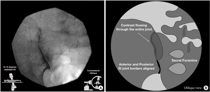
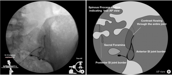
| 08:51, 15 April 2022 | SIJ fluoroscopy AP technique.jpg (file) |  |
40 KB | Jeremy | Anteroposterior technique fluoroscopic view (A) and graphical illustration (B). In the anteriorposterior (AP) approach, image is taken with 5–15 degree cephalad tilt from the vertical of the fluoroscopy machine reveals a joint with 2 separately visible anterior and posterior joint lines. The anterior and posterior parts of the sacroiliac (SI) joint were delineated as lateral and medial joint spaces, respectively. From Chauhan G, Hehar P, Loomba V, Upadhyay A. A Randomized Controlled Trial of... | 1 |
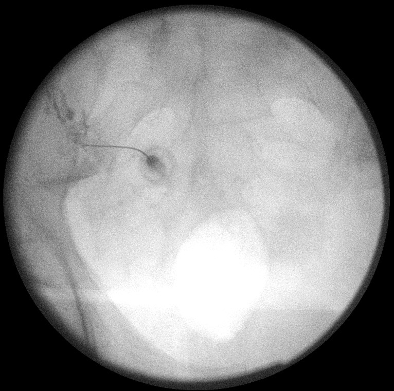
| 08:22, 15 April 2022 | SIJ injection fluoroscopy.jpg (file) |  |
141 KB | Jeremy | 1 | |
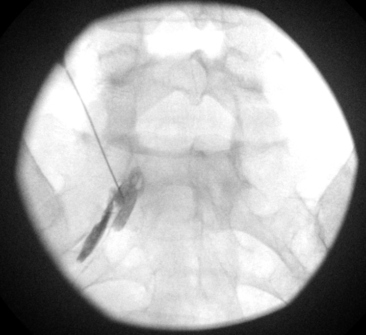
| 08:12, 15 April 2022 | S1 transforaminal injection fluoroscopy.jpg (file) | 88 KB | Jeremy | 1 | ||
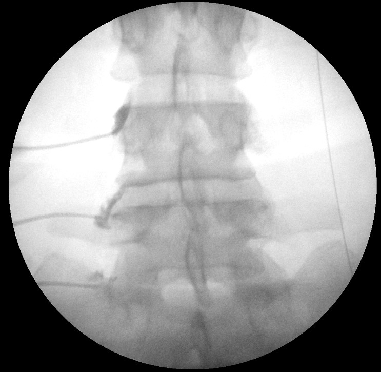
| 20:43, 13 April 2022 | Lumbar medial branch blocks fluoroscopy left L3-5.jpg (file) |  |
81 KB | Jeremy | Right L3 + L4 medial branch and L5 dorsal ramus blocks | 1 |
| 10:51, 13 April 2022 | Autonomic Dysfunction - Wells 2016.pdf (file) | 97 KB | Jeremy | Uploaded with SimpleBatchUpload | 1 | |
| 08:07, 13 April 2022 | Topical GTN for tendinopathies systematic review - Challoumas 2019.pdf (file) | 864 KB | Jeremy | 1 | ||
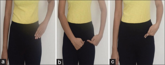
| 21:30, 12 April 2022 | Hip pain pointing.jpg (file) |  |
26 KB | Jeremy | Patient finger pointing indicative of hip joint pain. (a) Trochanteric C sign, (b) triangular sign, and (c) deep pointer sign From https://www.ncbi.nlm.nih.gov/pmc/articles/PMC8022067/ | 1 |
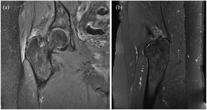
| 20:03, 11 April 2022 | Gluteus medius MRI high grade partial thickness tear.jpg (file) |  |
85 KB | Jeremy | (a) Coronal fat suppressed proton density and (b) sagittal T2-weighted sequences on MRI of the right hip showing a high-grade partial tear of the gluteus medius and minimus tendons with tendinosis and underlying trochanteric bursitis. The patient consented for publication of this imaging. Pianka MA, Serino J, DeFroda SF, Bodendorfer BM. Greater trochanteric pain syndrome: Evaluation and management of a wide spectrum of pathology. SAGE Open Med. 2021 Jun 3;9:20503121211022582. doi: 10.1177/20... | 1 |
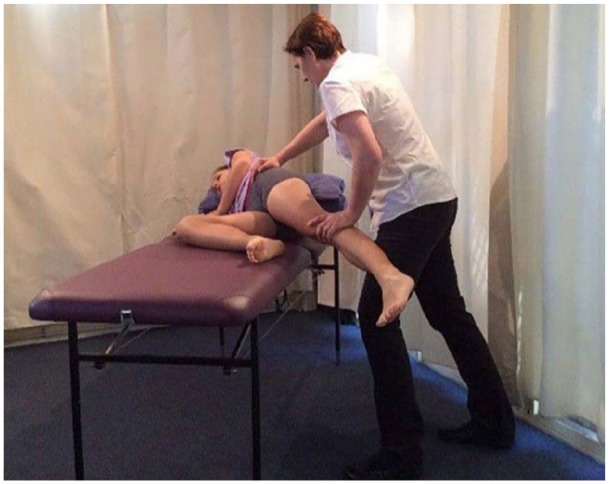
| 19:57, 11 April 2022 | Hip abductor strength testing.jpg (file) |  |
96 KB | Jeremy | Evaluation of hip abductor strength. The patient lies in the lateral decubitus position with the affected side facing up. With the hip and knee extended, the examiner asks the patient to abduct the hip against resistance Pianka MA, Serino J, DeFroda SF, Bodendorfer BM. Greater trochanteric pain syndrome: Evaluation and management of a wide spectrum of pathology. SAGE Open Med. 2021 Jun 3;9:20503121211022582. doi: 10.1177/20503121211022582. PMID: 34158938; PMCID: PMC8182177. | 1 |
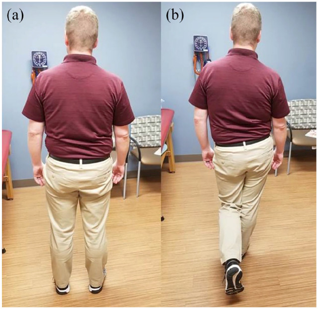
| 19:57, 11 April 2022 | Trendelenburg test.jpg (file) |  |
123 KB | Jeremy | Trendelenburg test. From a (a) standing position, (b) the patient is asked to stand on the affected leg and lift the contralateral foot off the ground. The test is considered positive, if the contralateral pelvis tilts downward, indicating abductor weakness Pianka MA, Serino J, DeFroda SF, Bodendorfer BM. Greater trochanteric pain syndrome: Evaluation and management of a wide spectrum of pathology. SAGE Open Med. 2021 Jun 3;9:20503121211022582. doi: 10.1177/20503121211022582. PMID: 34158938;... | 1 |

