Search results
From WikiMSK
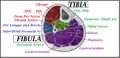
File:Leg compartments.jpg https://orthopaedia.com/page/Tibia-Fractures This work is licensed under the Creative Commons Attribution-NonCommercial-ShareAlike License.(946 × 461 (93 KB)) - 18:33, 13 March 2023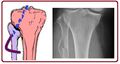
File:Peroneal nerve course.jpg https://orthopaedia.com/page/Tibia-Fractures This work is licensed under the Creative Commons Attribution-NonCommercial-ShareAlike License.(902 × 482 (48 KB)) - 18:33, 13 March 2023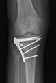
File:Tibial plateau ORIF.jpg https://orthopaedia.com/page/Tibia-Fractures This work is licensed under the Creative Commons Attribution-NonCommercial-ShareAlike License.(398 × 584 (18 KB)) - 18:34, 13 March 2023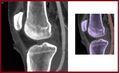
File:Popliteal artery knee xr.jpg https://orthopaedia.com/page/Tibia-Fractures This work is licensed under the Creative Commons Attribution-NonCommercial-ShareAlike License.(761 × 463 (34 KB)) - 18:33, 13 March 2023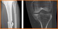
File:Tibial shaft comminuted fracture.jpg https://orthopaedia.com/page/Tibia-Fractures This work is licensed under the Creative Commons Attribution-NonCommercial-ShareAlike License.(936 × 478 (51 KB)) - 18:34, 13 March 2023- fractures, caused by excessive dorsiflexion of the foot against the distal tibia, comprise half of all talus fractures (Figure 6). They are classified, with19 KB (2,757 words) - 06:02, 2 April 2022
- fractures in the lower limb are commonly observed in various locations: tibia (23%), fibula (15%), tarsal navicular (17%), and metatarsals (16%). They17 KB (2,615 words) - 19:09, 11 November 2023
- Distal Phalanx (Foot) Knee Joint Joint type: Saddle Joint, Hinge Joint Bones: Tibia, Femur, Patella Innervation: Popliteal nerve, tibial nerve Vasculature: Genicular26 members (1 subcategory, 0 files) - 20:19, 1 May 2022


