Search results
From WikiMSK

File:Anatomy.png (128 × 128 (5 KB)) - 16:55, 3 March 2022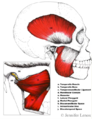
File:TMJ Anatomy.png File uploaded with MsUpload(518 × 672 (377 KB)) - 09:15, 28 June 2020
File:Z anatomy screenshot.jpg (1,920 × 1,080 (231 KB)) - 21:11, 6 November 2022
File:Head anatomy drawing.png (225 × 256 (83 KB)) - 08:25, 6 April 2021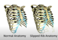
File:Slipped rib anatomy.png File uploaded with MsUpload(607 × 419 (277 KB)) - 06:41, 2 February 2021
File:Cauda equina anatomy.jpg File uploaded with MsUpload(229 × 468 (26 KB)) - 19:57, 16 May 2021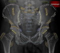
File:Pelvic Xray Anatomy.PNG File uploaded with MsUpload(596 × 527 (521 KB)) - 19:02, 30 August 2020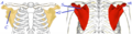
File:Scapula anatomy.png https://orthopaedia.com/page/Scapular-Fractures This work is licensed under a Creative Commons Attribution-NonCommercial-NoDerivatives 4.0 International(624 × 160 (121 KB)) - 20:58, 11 March 2023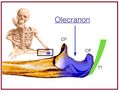
File:Olecranon anatomy.jpg https://orthopaedia.com/page/Olecranon-Fractures This work is licensed under the Creative Commons Attribution-NonCommercial-ShareAlike License.(675 × 518 (43 KB)) - 21:08, 11 March 2023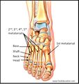
File:Metatarsal anatomy.jpg From https://orthopaedia.com/page/Metatarsal-Fractures This work is licensed under the Creative Commons Attribution-NonCommercial-ShareAlike License.(805 × 847 (82 KB)) - 07:04, 8 March 2022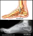
File:Hindfoot anatomy.jpg From https://orthopaedia.com/page/Hindfoot-Fractures This work is licensed under the Creative Commons Attribution-NonCommercial-ShareAlike License.(824 × 900 (88 KB)) - 09:12, 8 March 2022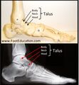
File:Talar anatomy.jpg From https://orthopaedia.com/page/Hindfoot-Fractures This work is licensed under the Creative Commons Attribution-NonCommercial-ShareAlike License.(834 × 900 (89 KB)) - 09:22, 8 March 2022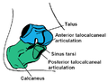
File:Subtalar joint anatomy basic.png File uploaded with MsUpload(724 × 558 (24 KB)) - 13:22, 17 July 2021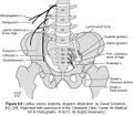
File:Lumbar-Plexus-Anatomy-1.jpg File uploaded with MsUpload(600 × 526 (101 KB)) - 12:36, 16 April 2021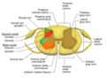
File:Spainal cord sectional anatomy.png File uploaded with MsUpload(1,200 × 880 (124 KB)) - 09:12, 17 May 2021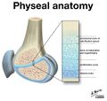
File:Physeal anatomy.jpg Figure 2: Physeal anatomy showing the four zones. (Case courtesy of Dr Matt Skalski, Radiopaedia.org. From the case https://radiopaedia.org/cases/27354)(1,600 × 1,516 (146 KB)) - 19:31, 8 March 2022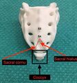
File:Sacral anatomy.jpg From https://resources.wfsahq.org/atotw/ultrasound-guided-caudal-anaesthesia/ This work is licensed under a Creative Commons Attribution-NonCommercial-NoDerivatives(680 × 726 (101 KB)) - 12:18, 30 April 2022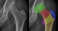
File:Hip xr anatomy.jpg https://orthopaedia.com/page/Hip-Fractures This work is licensed under the Creative Commons Attribution-NonCommercial-ShareAlike License.(1,957 × 1,053 (208 KB)) - 05:16, 13 March 2023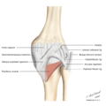
File:Posterior knee anatomy.png Case courtesy of Assoc Prof Frank Gaillard, Radiopaedia.org. From the case rID: 9330 This work is licensed under the Creative Commons Attribution-NonC(1,200 × 1,200 (172 KB)) - 11:18, 3 August 2021
File:Ankle us anatomy.jpg From https://wikem.org/wiki/File:Ankle_us_anatomy.png This work is licensed under the Creative Commons Attribution-ShareAlike 4.0 International License(1,118 × 856 (59 KB)) - 18:35, 5 May 2022


