Search results
From WikiMSK
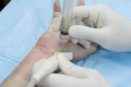
File:TMC joint ultrasound transducer placement umphrey.png Ultrasound transducer over TMC joint. Needle is positioned on the volar side of the transducer at the midpoint of the transducer’s long axis. Path of entry(498 × 330 (230 KB)) - 07:31, 15 June 2021
File:Sacroiliac Joint and Facet Joint Diagnostic Tests Review - Hancock 2007.pdf File uploaded with MsUpload(261 KB) - 19:21, 18 May 2021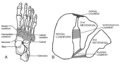
File:Lisfranc joint.png File uploaded with MsUpload(1,168 × 614 (256 KB)) - 10:13, 21 June 2020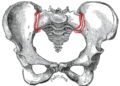
File:SI joint.png File uploaded with MsUpload(450 × 321 (131 KB)) - 10:01, 23 August 2020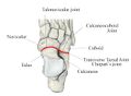
File:Transverse-tarsal-joint.jpg File uploaded with MsUpload(659 × 481 (30 KB)) - 18:50, 17 July 2021
File:MBB facet joint.png File uploaded with MsUpload(403 × 397 (31 KB)) - 20:38, 11 July 2020
File:Posterior sacroiliac joint.png File uploaded with MsUpload(559 × 700 (95 KB)) - 22:35, 3 August 2020
File:Anterior sacroiliac joint.png File uploaded with MsUpload(526 × 677 (98 KB)) - 22:35, 3 August 2020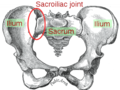
File:Sacroiliac joint Gray.png File uploaded with MsUpload(300 × 225 (103 KB)) - 13:38, 19 April 2021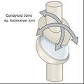
File:Condyloid joint.jpg From OpenStax college This work is licensed under a Creative Commons Attribution 4.0 International License.(737 × 737 (144 KB)) - 06:48, 2 April 2022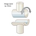
File:Hinge joint.jpg From OpenStax college This work is licensed under a Creative Commons Attribution 4.0 International License.(704 × 704 (105 KB)) - 06:49, 2 April 2022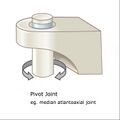
File:Pivot joint.jpg From OpenStax college This work is licensed under a Creative Commons Attribution 4.0 International License.(687 × 687 (101 KB)) - 06:50, 2 April 2022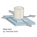
File:Plane joint.jpg From OpenStax college This work is licensed under a Creative Commons Attribution 4.0 International License.(700 × 700 (108 KB)) - 06:50, 2 April 2022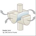
File:Saddle joint.jpg From OpenStax college This work is licensed under a Creative Commons Attribution 4.0 International License.(798 × 798 (157 KB)) - 06:50, 2 April 2022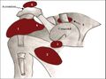
File:Bursae shoulder joint.jpg Diagram of normal bursae surrounding the shoulder joint: (1) subacromial-subdeltoid bursa, (2) subscapular recess, (3) subcoracoid bursa, (4) coracoclavicular(730 × 544 (103 KB)) - 17:17, 16 August 2021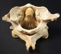
File:Atlantoaxial joint.jpg https://commons.wikimedia.org/wiki/File:Vertebra_-_atlas,_axis_(superior).jpg This work is licensed under the Creative Commons Zero Public Domain License(1,002 × 887 (131 KB)) - 14:53, 2 April 2022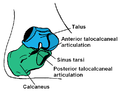
File:Subtalar joint anatomy basic.png File uploaded with MsUpload(724 × 558 (24 KB)) - 13:22, 17 July 2021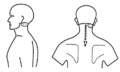
File:Atlanto-axial joint pattern dreyfuss.png Copyrighted. Dreyfuss et al.. Atlanto-occipital and lateral atlanto-axial joint pain patterns. Spine 1994. 19:1125-31. PMID: 8059267. DOI.(722 × 436 (22 KB)) - 18:35, 23 May 2021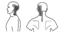
File:Atlanto-occipital joint pattern dreyfuss.png Copyrighted. Dreyfuss et al.. Atlanto-occipital and lateral atlanto-axial joint pain patterns. Spine 1994. 19:1125-31. PMID: 8059267. DOI.(480 × 277 (30 KB)) - 18:34, 23 May 2021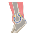
File:Elbow joint.png https://commons.wikimedia.org/wiki/File:202107_Sagittal_section_through_the_elbow_joint.svg This work is licensed under the Creative Commons Attribution-ShareAlike(400 × 400 (39 KB)) - 07:25, 2 April 2022


