Search results
From WikiMSK

File:Degenerative Joint Disease of the Spine - Bogduk 2012.pdf File uploaded with MsUpload(510 KB) - 19:45, 14 August 2021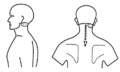
File:Atlanto-axial joint pattern dreyfuss.png Dreyfuss et al.. Atlanto-occipital and lateral atlanto-axial joint pain patterns. Spine 1994. 19:1125-31. PMID: 8059267. DOI.(722 × 436 (22 KB)) - 18:35, 23 May 2021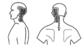
File:Atlanto-occipital joint pattern dreyfuss.png Dreyfuss et al.. Atlanto-occipital and lateral atlanto-axial joint pain patterns. Spine 1994. 19:1125-31. PMID: 8059267. DOI.(480 × 277 (30 KB)) - 18:34, 23 May 2021
File:IDD pic.jpg Classification of internal disc disruption Copyright 2012 Published by Elsevier Inc Bogduk N. Degenerative joint disease of the spine. Radiol Clin North(316 × 279 (22 KB)) - 18:34, 15 August 2021
File:Aizawa sections.PNG location depends on the affected section of the sacroiliac joint. European spine journal : official publication of the European Spine Society, the European Spinal(582 × 596 (252 KB)) - 19:19, 1 September 2020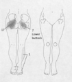
File:Aizawa section 3.png location depends on the affected section of the sacroiliac joint. European spine journal : official publication of the European Spine Society, the European Spinal(485 × 555 (271 KB)) - 19:19, 1 September 2020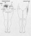
File:Aizawa section 0.png location depends on the affected section of the sacroiliac joint. European spine journal : official publication of the European Spine Society, the European Spinal(483 × 555 (281 KB)) - 19:19, 1 September 2020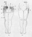
File:Aizawa section 1.png location depends on the affected section of the sacroiliac joint. European spine journal : official publication of the European Spine Society, the European Spinal(485 × 555 (281 KB)) - 19:19, 1 September 2020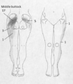
File:Aizawa section 2.png location depends on the affected section of the sacroiliac joint. European spine journal : official publication of the European Spine Society, the European Spinal(485 × 555 (278 KB)) - 19:19, 1 September 2020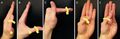
File:Motor actions of hand musles innervated by median nerve.jpg neuropathy: a survey of 24 spine surgeons. Global Spine J. 2014 Feb;4(1):1-6. doi: 10.1055/s-0033-1354254. Epub 2013 Aug 28. PMID: 24494175; PMCID: PMC3908974(800 × 262 (169 KB)) - 09:20, 23 April 2022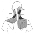
File:Cervical facet joint pain map.png maps with noxious stimulation of cervical facet joints in volunteers. Copyrighted Dwyer et al.. Cervical zygapophyseal joint pain patterns. I: A study in(450 × 454 (105 KB)) - 11:54, 23 May 2021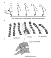
File:Whiplash cervical spine motion.png phase. C: The hypothesis of the whiplash injury mechanism. In the S-shaped position, the motion segment at the apex of the convex curvature receives rotational(463 × 524 (99 KB)) - 17:45, 23 May 2021File:Sacroiliac joint ultrasound cineloop.mp4 Sacroiliac joint ultrasound cineloop. Starting transverse plane from lower lumbar spine, moving caudally, to S2, then laterally to the sacroiliac joint. This(2.6 MB) - 08:18, 5 August 2020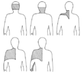
File:Cervical disc pain joint map Grubb.png is reproduced in a limited way under the fair-use doctrine. It falls under the "Non-profit educational" clause of the Fair Use doctrine.(524 × 469 (51 KB)) - 18:38, 23 May 2021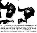
File:Cervical Rotation Lateral Flexion Test.jpg moving the ear towards the chest. The test is positive if a bony restriction totally blocked the lateral flexion part of the movement. Subluxation of the(536 × 481 (77 KB)) - 22:17, 18 February 2022


