Search results
From WikiMSK
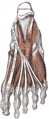
File:Muscles plantar foot Gray444.png This work is part of the public domain.(330 × 800 (59 KB)) - 11:10, 16 April 2022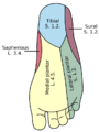
File:Gray834 plantar foot sensation.png This work is part of the public domain.(578 × 768 (69 KB)) - 21:53, 4 May 2022
File:Plantar Fibromatosis Review - Carroll 2018.pdf File uploaded with MsUpload(674 KB) - 18:54, 20 February 2024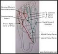
File:Mortons neuroma plantar nerves.jpg This work is licensed under the Creative Commons Attribution-NonCommercial-ShareAlike License.(957 × 892 (93 KB)) - 05:00, 7 May 2022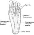
File:Plantar fascia.jpg From https://upload.wikimedia.org/wikipedia/commons/b/b1/PF-PlantarDesignCrop.jpg This work is licensed under the Creative Commons Attribution-ShareAlike(771 × 786 (69 KB)) - 21:50, 15 April 2022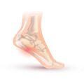
File:Plantar fasciitis.jpg From https://upload.wikimedia.org/wikipedia/commons/1/1a/Arch_tendonitis.jpg This work is licensed under the Creative Commons Attribution-ShareAlike 4(2,000 × 1,996 (156 KB)) - 21:53, 15 April 2022
File:Muscles plantar foot first layer Sobo.jpg This work is part of the public domain.(719 × 1,010 (110 KB)) - 11:12, 16 April 2022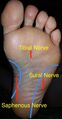
File:Plantar Nerve Distribution.jpg From https://wikem.org/wiki/File:Plantar_Nerve_Distribution.jpg This work is licensed under the Creative Commons Attribution-ShareAlike 4.0 International(469 × 898 (48 KB)) - 21:54, 4 May 2022
File:Plantar Foot Nerve Distribution.jpg From https://wikem.org/wiki/File:Plantar_Foot_Nerve_Distribution.jpg This work is licensed under the Creative Commons Attribution-ShareAlike 4.0 International(438 × 899 (46 KB)) - 21:55, 4 May 2022File:Evaluation and Treatment of Chronic Plantar Fasciitis - Latt 2020.pdf File uploaded with MsUpload(819 KB) - 21:52, 15 April 2022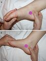
File:Plantar flexion inversion and dorsiflexion and eversion tarsal tunnel.jpg Reproduced with permission from A.M. Trescot (ed.), Peripheral Nerve Entrapments: Clinical Diagnosis and Management, DOI 10.1007/978-3-319-27482-9_74 This(350 × 456 (32 KB)) - 19:24, 16 April 2022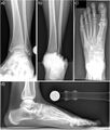
File:Foot and Ankle Radiograph Normal.jpg and ankle: a) anteroposterior view; b) hindfoot alignment view; c) dorso-plantar view; d) lateral view. Krähenbühl, Nicola et al. “The subtalar joint: A(647 × 761 (59 KB)) - 13:51, 17 July 2021


