Uncategorised files
From WikiMSK
Showing below up to 100 results in range #201 to #300.
 Closed access.png 576 × 900; 21 KB
 Coccyx imaging - Skalski 2020.pdf ; 3.72 MB
 Concepts.png 256 × 256; 21 KB
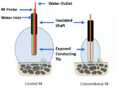 Cooled vs Conventional RF.png 529 × 389; 91 KB
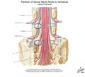
 Coronal spinal nerve relationship.jpg 1,159 × 768; 132 KB
 Corticospinal Tracts.jpg 582 × 1,273; 166 KB
- Corticosteroid Injections - 2015.pdf ; 128 KB
- Corticosteroids Structure Acty Relns.pdf ; 1.64 MB
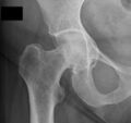 Coxa profunda xray.jpg 523 × 491; 30 KB
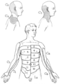
 Cranial dermatomes.png 1,029 × 495; 213 KB
 Creative commons licenses comparisons.jpg 800 × 555; 71 KB
 Creep.jpg 400 × 222; 8 KB
- Critcal Overview MPS 2019.pdf ; 289 KB
 Crossover posterior wall and ischial spine signs xray.png 426 × 277; 32 KB
 DDx.png 256 × 256; 14 KB
- DNS Exercises - Kolar 2015.pdf ; 2.99 MB
- DNS Reflex Stimulation - Kolar 2007.pdf ; 1.51 MB
 Davis cervical dermatomes.png 329 × 323; 11 KB
 Davis lumbar dermatomes.png 468 × 356; 20 KB
 De Quervain Compartments.PNG 309 × 583; 178 KB
 De Quervain Ultrasound Injection.PNG 378 × 274; 160 KB
- Deep Somatic Pain - NB.pdf ; 1.28 MB
 Definition.png 256 × 256; 15 KB
 Dermatome Types.png 3,550 × 2,377; 186 KB
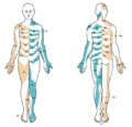
 Dermatome map Keegan and garrett.png 800 × 866; 776 KB
 Dermatome map head and campbell.jpeg 767 × 732; 134 KB
 Dermatome map herringham.jpeg 273 × 809; 22 KB
 Dermatome map lee.PNG 677 × 649; 320 KB
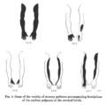
 Dermatome map lower body Fender after Foerster.png 800 × 1,164; 211 KB
 Dermatome map sherrington.jpeg 693 × 636; 149 KB
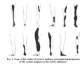
 Dermatome map upper body Fender after Foerster.png 800 × 1,106; 216 KB
 Dermatomyositis.jpg 768 × 656; 80 KB
 Determinants of disc degeneration.PNG 502 × 390; 60 KB
 Diagnostic accuracy table.png 794 × 232; 82 KB
 Diagnostic accuracy table example.png 1,028 × 188; 98 KB
 Diagnostic criteria summary.PNG 696 × 738; 86 KB
 Diagram-classification-of-acl-avulsion-fractures-1.jpg 900 × 900; 199 KB
 Differential lateral elbow tendinopathy.PNG 1,057 × 752; 182 KB
 Disc innervation.jpg 700 × 454; 59 KB
 Disc protrusion posterior view.jpg 842 × 767; 80 KB
 Doctor.jpg 332 × 408; 39 KB
 Doctorf.png 512 × 512; 27 KB
 Doctorm.png 512 × 512; 28 KB
 Donation.png 256 × 256; 11 KB
 Dorsal-wrist-pain.jpg 433 × 647; 55 KB
 Dorsal rami branches.PNG 359 × 328; 29 KB
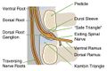
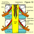 Dural sleeve.png 400 × 400; 161 KB
- EBM Acute Musculoskeletal Pain.pdf ; 1.29 MB
 EBQs.png 256 × 256; 4 KB
- ESSR ankle.pdf ; 2.82 MB
- ESSR elbow.pdf ; 3.4 MB
- ESSR hip.pdf ; 3.29 MB
- ESSR knee.pdf ; 1.34 MB


