Uncategorised files
From WikiMSK
Showing below up to 250 results in range #251 to #500.
 Dermatome Types.png 3,550 × 2,377; 186 KB
 Dermatome map Keegan and garrett.png 800 × 866; 776 KB
 Dermatome map head and campbell.jpeg 767 × 732; 134 KB
 Dermatome map herringham.jpeg 273 × 809; 22 KB
 Dermatome map lee.PNG 677 × 649; 320 KB
 Dermatome map lower body Fender after Foerster.png 800 × 1,164; 211 KB
 Dermatome map sherrington.jpeg 693 × 636; 149 KB
 Dermatome map upper body Fender after Foerster.png 800 × 1,106; 216 KB
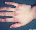 Dermatomyositis.jpg 768 × 656; 80 KB
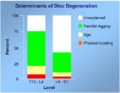 Determinants of disc degeneration.PNG 502 × 390; 60 KB
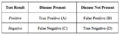 Diagnostic accuracy table.png 794 × 232; 82 KB
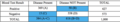 Diagnostic accuracy table example.png 1,028 × 188; 98 KB
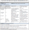 Diagnostic criteria summary.PNG 696 × 738; 86 KB
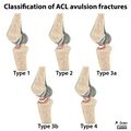 Diagram-classification-of-acl-avulsion-fractures-1.jpg 900 × 900; 199 KB
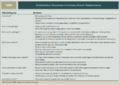 Differential lateral elbow tendinopathy.PNG 1,057 × 752; 182 KB
 Disc innervation.jpg 700 × 454; 59 KB
 Disc protrusion posterior view.jpg 842 × 767; 80 KB
 Doctor.jpg 332 × 408; 39 KB
 Doctorf.png 512 × 512; 27 KB
 Doctorm.png 512 × 512; 28 KB
 Donation.png 256 × 256; 11 KB
 Dorsal-wrist-pain.jpg 433 × 647; 55 KB
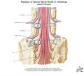
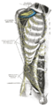
 Dorsal rami branches.PNG 359 × 328; 29 KB
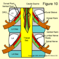 Dural sleeve.png 400 × 400; 161 KB
 EBM Acute Musculoskeletal Pain.pdf ; 1.29 MB
 EBQs.png 256 × 256; 4 KB
- ESSR ankle.pdf ; 2.82 MB
- ESSR elbow.pdf ; 3.4 MB
- ESSR hip.pdf ; 3.29 MB
- ESSR knee.pdf ; 1.34 MB
- ESSR shoulder.pdf ; 2.54 MB
- ESSR wrist.pdf ; 2.7 MB
 Edit.png 128 × 128; 5 KB
 EditToolbar2.png 1,148 × 138; 30 KB
 Elbow.png 128 × 128; 5 KB
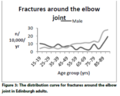 Elbow fractures incidence.png 360 × 287; 32 KB
 Electrodx Periph Nerve.doc ; 40 KB
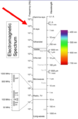 Electromagnetic spectrum.PNG 375 × 574; 65 KB
 Ellerslie Medical Centre.png 455 × 167; 19 KB
- Epid Nat Hx Risk Factors.doc ; 46 KB
- Erythromelalgia - Mann 2018.pdf ; 171 KB
 Exam.png 256 × 256; 9 KB
 Examination.png 128 × 128; 7 KB
- Exercise Prescription - Luan 2019.pdf ; 1.09 MB
 Exercise induced pain and analgesia mechanisms.jpeg 650 × 928; 128 KB
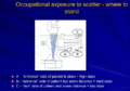 Exposure doses scatter.PNG 717 × 500; 230 KB
 External-links.png 256 × 256; 9 KB
 F1.large.jpg 2,000 × 2,000; 455 KB
- Fabry Disease - ortiz 2018.pdf ; 424 KB
- Fabry disease - Germain 2010.pdf ; 2.21 MB
 Fair use logo.svg.png 768 × 768; 21 KB
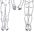
 Falconer L5 dermatome.png 405 × 450; 21 KB
 Falconer S1 dermatome.png 392 × 442; 24 KB
- Fascia review - wilke 2017.pdf ; 992 KB
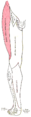
 Fear Avoidance Model.jpg 1,237 × 728; 94 KB
 Feedback.png 256 × 256; 8 KB
 Femoral nerve.jpeg 630 × 630; 50 KB
- Fibromyalgia Review - Clauw 2014.pdf ; 406 KB
- Fibromyalgia year in review 2020.pdf ; 239 KB
- Fibromyalgia year in review 2021.pdf ; 381 KB
- Fibromyalgia year in review 2022.pdf ; 366 KB
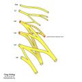
 Finger annular ligaments.png 498 × 478; 57 KB
 First Dorsal Compartment.PNG 431 × 278; 189 KB
 Flexor retinaculum wrist.png 2,860 × 1,031; 35 KB
 Fluoroscopy system.PNG 552 × 365; 25 KB
 Foerster dermatomes redrawn by Lee.png 726 × 716; 83 KB
 Foot.png 128 × 128; 5 KB
- Fullerton2018.pdf ; 2.49 MB
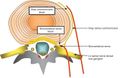
 GON schematic.PNG 292 × 180; 16 KB
- GRASP protocol.pdf ; 676 KB
 Gabapentinoids vs placebo adverse effects infographic Mathieson.png 1,453 × 622; 147 KB
 Gabapentinoids vs placebo pain infographic Mathieson.png 1,488 × 638; 168 KB
 Genicular.jpg 500 × 427; 603 KB
 Genitofemoral nerve.jpeg 630 × 630; 48 KB
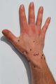
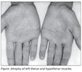 Gillat-Sumner hand.jpg 360 × 316; 19 KB
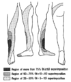
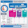 Gluteal tendinopathy va v12.png 1,024 × 1,024; 127 KB
 Gray337 Metacarpophalangeal joint and digit palmar.png 235 × 550; 18 KB
 Gray338 Metacarpophalangeal joint and digit ulnar.png 283 × 550; 15 KB
 Greater-and-lesser-occipital-nerve-blocks.png 423 × 516; 223 KB
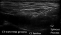
 Greater occipital nerve block ultrasound.jpg 960 × 444; 42 KB
 Greater occipital nerve injection.PNG 375 × 220; 63 KB
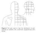 Grids.jpg 1,000 × 926; 106 KB
- Grimaldi2015 - Gluteal Tendinopathy.pdf ; 2.94 MB
 Guidelines.png 128 × 128; 3 KB
- HISTAMINE 2.doc ; 43 KB
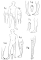
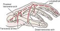 Hand arches.jpg 491 × 254; 32 KB
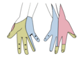 Handnerves.png 355 × 252; 31 KB
- Harden2010 - Budapest Criteria.pdf ; 148 KB
 Haymaker and Woodhall.png 417 × 500; 67 KB
 Head.png 128 × 128; 6 KB
 Head anatomy drawing.png 225 × 256; 83 KB
 Head dermatomes redrawn by Lee.png 720 × 679; 72 KB
 Henderson-Hasselbalch-equation-for-drug-dissociation.jpg 802 × 176; 21 KB
 Hip.png 128 × 128; 4 KB
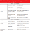 Hip Nerve Entrapment Table.PNG 434 × 445; 66 KB
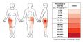 Hip OA pain distribution.jpg 1,427 × 741; 142 KB
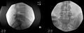
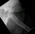
- Error creating thumbnail: Unable to save thumbnail to destinationHip fluoroscopy.jpg 1,241 × 1,210; 124 KB
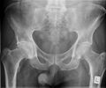 Hip x-ray frontal advanced osteoarthritis.jpeg 836 × 694; 155 KB
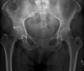 Hip x-ray frontal early osteoarthritis.png 760 × 644; 180 KB
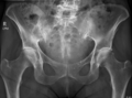 Hip x-ray frontal moderate osteoarthritis.png 882 × 656; 261 KB
 Holtzhausen.jpg 480 × 581; 30 KB
 Human-bones.png 128 × 128; 10 KB
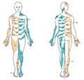
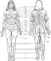
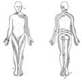
 Human skeleton front2.png 310 × 599; 100 KB
 Hyperalgesia and allodynia.png 3,499 × 1,841; 42 KB
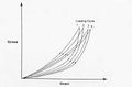 Hysteresis.jpg 400 × 262; 19 KB
- IASP Taxonomy 1994.pdf ; 1.95 MB
 IDD pic.jpg 316 × 279; 22 KB
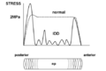 IDD vs normal disc stress profilometry.png 532 × 360; 52 KB
 IPM journal.jpg 480 × 664; 48 KB
 ISO pain case 0.png 475 × 744; 162 KB
 ISO pain case 0 post injection.png 422 × 744; 122 KB
 ISO pain case 1.png 189 × 322; 78 KB
 ISO pain case 2.png 224 × 247; 83 KB
 ISO pain case 3.png 178 × 322; 86 KB
 ISO pain case 4.png 433 × 398; 161 KB
 ISO pain case 5.png 253 × 260; 88 KB
 Iliohypogastric nerve.jpeg 630 × 630; 48 KB
 Ilioinguinal nerve.jpeg 630 × 630; 48 KB
 Important.png 128 × 128; 6 KB
 Incidence OA hand hip and knee.png 424 × 418; 65 KB
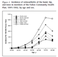
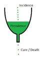 Incidence Prevalence.jpg 372 × 531; 20 KB
- Inclusion Body Myositis - Goyal 2022.pdf ; 5.18 MB
 Inferior gluteal nerve.jpeg 630 × 630; 55 KB
 Inflammation.png 128 × 128; 7 KB
- Injection Pain Assessment Form.pdf ; 127 KB
- Injection Pain Form.pdf ; 103 KB
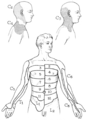
 Intercostal nerve Gray.png 473 × 400; 21 KB
 Intercostal nerves anterior view Gray.png 358 × 700; 66 KB
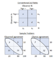 Interobserver Reliability and the Kappa Statistic.png 400 × 430; 18 KB
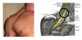 Interscalene block.jpg 1,200 × 590; 277 KB
 Interspinous Oedema Fluoroscopic Injection.jpg 938 × 378; 87 KB
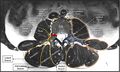
 Interspinous Oedema MRI.jpg 930 × 433; 80 KB
 Interspinous Oedema MRI2.png 832 × 508; 222 KB
 Jenny K.jpg 660 × 876; 73 KB
 Jeremy S.jpg 852 × 852; 129 KB
 Journal osteopathic medicine.jpg 800 × 1,066; 54 KB
 Kallikrein kinin system.png 2,303 × 1,791; 54 KB
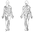
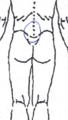
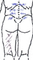
 Keegan and Garrett.png 403 × 500; 57 KB
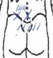
 Keegan and Garrett dermatome embryology theory.png 514 × 482; 27 KB
 Keegan and Garrett dermatomes redrawn by Lee.png 674 × 662; 66 KB
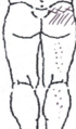
 Keegan and Garrett lumbar dermatomes.png 611 × 681; 108 KB
 Keegan and Garrett original map.jpg 682 × 699; 111 KB
 Kellgren cervical pain.PNG 706 × 671; 156 KB
 Kellgren lumbar pain.PNG 733 × 443; 142 KB
 Kellgren thoracic pain.PNG 724 × 459; 102 KB
- Khan tendonosis report.pdf ; 357 KB
 Knee.png 128 × 128; 6 KB
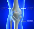
 Knee diagram2.png 658 × 600; 238 KB
 Knee surgery Skou et al.png 521 × 443; 54 KB
 L4-S1 nerve block band like Nitta.png 472 × 527; 45 KB
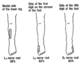 L4-S1 nerve block distinctive sensory deficit Nitta.png 408 × 319; 29 KB
 L4 axial spinal nerves.jpg 750 × 451; 47 KB
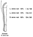
 L4 nerve block sensory deficit Nitta.png 417 × 480; 75 KB
 L5 nerve block sensory deficit Nitta.png 405 × 479; 72 KB
- LEAP Protocol.pdf ; 1.34 MB
 LFCN schematic.jpg 600 × 1,202; 88 KB
 LFCN ultrasound.jpg 600 × 452; 71 KB
 LFCN ultrasound2.jpg 600 × 454; 70 KB
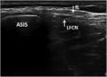 LFCN ultrasound3.jpg 600 × 428; 57 KB
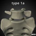 LSTV 1a.jpeg 1,024 × 1,024; 469 KB
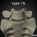 LSTV 1b.jpeg 1,024 × 1,024; 467 KB
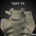 LSTV 2a.jpeg 1,024 × 1,024; 477 KB
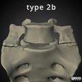 LSTV 2b.jpeg 1,024 × 1,024; 488 KB
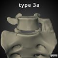 LSTV 3a.jpeg 1,024 × 1,024; 480 KB
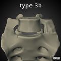 LSTV 3b.jpeg 1,024 × 1,024; 471 KB
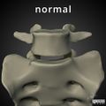 LSTV Normal.jpeg 1,024 × 1,024; 473 KB
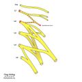
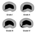
 LSTV Type 2a.jpg 410 × 298; 39 KB
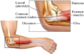
 Lateral Elbow Tendinopathy.png 600 × 600; 312 KB
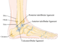 Lateral collateral ligament of ankle.png 1,830 × 1,309; 146 KB
 Lateral elbow.png 488 × 318; 213 KB
 Lateral femoral cutaneous nerve.jpeg 630 × 630; 49 KB
 Lateral femoral cutaneous nerve skin innervation.png 239 × 900; 20 KB
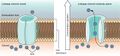 Leakage-channel.jpg 1,119 × 525; 129 KB
 Leg pain case 001 L hip lateral.png 563 × 534; 88 KB
 Leg pain case 001 MRI Lumbar L4-5 Transarticular.jpg 1,084 × 1,020; 83 KB
 Leg pain case 001 MRI Lumbar Spine Sagittal Median.jpg 570 × 1,108; 81 KB
 Leg pain case 001 MRI Lumbar Spine Sagittal Paramedian.jpg 632 × 1,108; 90 KB
 Leg pain case 001 MRI angiogram.png 558 × 601; 136 KB
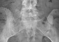
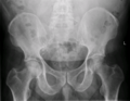 Leg pain case 001 pelvis AP.png 847 × 659; 257 KB
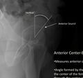 Lequesnes anterior centre edge angle ap film.jpg 215 × 203; 9 KB
 Leukocytes in PRP.jpg 922 × 692; 176 KB
 Library.png 60 × 62; 4 KB
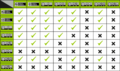 License Compatibility Chart.png 1,200 × 710; 220 KB
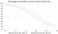 Lidocaine vs bupivicaine inactive form percentage vs ph.png 1,718 × 1,020; 108 KB


