Unused files
From WikiMSK
The following files exist but are not embedded in any page. Please note that other web sites may link to a file with a direct URL, and so may still be listed here despite being in active use.
Showing below up to 89 results in range #1 to #89.
- TMJ Opening MWM.mp4 ; 2.58 MB
- TMJ Closing MWM.mp4 ; 1.6 MB
- TMJ Lateral Deviation MWM.mp4 ; 2.56 MB
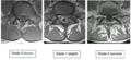
 C1-2.tif 475 × 446; 933 KB
- C2-3.tif 480 × 443; 880 KB
- C3-4.tif 487 × 430; 870 KB
- C4-5NL.tif 990 × 764; 763 KB
- C5-6.tif 478 × 425; 839 KB
- C6-7.tif 484 × 430; 872 KB
- Probability C1-C4.tif 504 × 461; 930 KB
- Probability C4-C7.tif 518 × 458; 964 KB
 Cervical Pain Maps Grid.jpg 2,065 × 3,019; 655 KB
 Cervical-Radicular-Pain-Patterns.pdf ; 1.06 MB
- NZAMM-Curriculum-FINAL-2019.pdf ; 951 KB
- NZAMM-Training-Manual-190118.pdf ; 628 KB
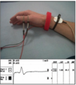
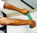 Rubber bar lateral elbow.jpg 292 × 266; 48 KB
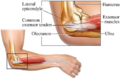 Lateral elbow.png 488 × 318; 213 KB
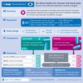 F1.large.jpg 2,000 × 2,000; 455 KB
- Bmjsem-2018-Holmich protocol.pdf ; 500 KB
 Hip Nerve Entrapment Table.PNG 434 × 445; 66 KB
 Nzamm-logo.png 274 × 109; 20 KB
 Yazbek progression program.PNG 807 × 791; 147 KB
- Injection Pain Assessment Form.pdf ; 127 KB
 Pubmed.png 16 × 16; 521 bytes
 Nzamm.png 113 × 109; 14 KB
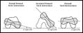
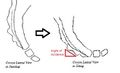 Coccyx angle of incidence.jpg 438 × 267; 32 KB
- Coccyx animation.webm ; 211 KB
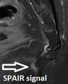 Coccyx distal spair.png 190 × 234; 46 KB
 Category section.PNG 1,367 × 392; 28 KB
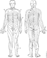
 Human skeleton front.png 310 × 599; 83 KB
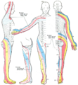 Dermatome map Keegan and garrett.png 800 × 866; 776 KB
 Pmc logo.png 38 × 20; 2 KB
 Open Access logo PLoS white.svg 640 × 1,000; 1 KB
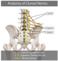 Cluneal nerves.PNG 485 × 510; 231 KB
 Caution.png 24 × 24; 485 bytes
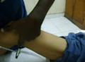 Medial hamstring reflex.PNG 502 × 369; 290 KB
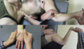 CTS manual therapy.png 557 × 325; 347 KB
 Radial nerve NCS SNAP.png 288 × 321; 135 KB
 Human skeleton front2.png 310 × 599; 100 KB
 Multifidus fat infiltration.png 671 × 307; 191 KB
 Head anatomy drawing.png 225 × 256; 83 KB
 Welcome to wikimsk.png 1,350 × 650; 301 KB
 WIKIMSK hero.png 1,300 × 350; 181 KB
 Red-flag2.png 32 × 32; 523 bytes
 Red-flag.png 32 × 32; 799 bytes
- Physiol deep somatic pain.pdf ; 387 KB
 Nzamm-logo-long.png 1,504 × 413; 59 KB
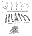 Whiplash cervical spine motion.png 463 × 524; 99 KB
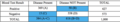 Diagnostic accuracy table example.png 1,028 × 188; 98 KB
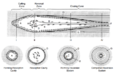 Cortical remodeling unit.png 1,003 × 607; 227 KB
 Keegan and Garrett.png 403 × 500; 57 KB
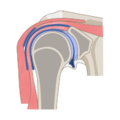 Coronal section of shoulder joint.png 400 × 400; 54 KB
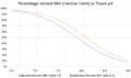 Lidocaine vs bupivicaine inactive form percentage vs ph.png 1,718 × 1,020; 108 KB
 Femoral torsion.jpeg 520 × 208; 17 KB
- NZCMM Training Manual Oct 2021.pdf ; 514 KB
 Median nerve position variations wrist.png 425 × 430; 96 KB
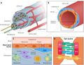 Blood nerve barrier.jpg 1,977 × 1,508; 263 KB
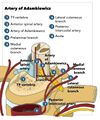 Artery of Adamkiewicz.jpg 753 × 901; 121 KB
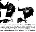 Cervical Rotation Lateral Flexion Test.jpg 536 × 481; 77 KB
 Anatomy.png 128 × 128; 5 KB
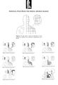
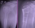 Proximal humeral fracture and fixation.jpg 962 × 768; 161 KB
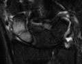 Occult scaphoid fracture MRI.jpg 300 × 236; 8 KB
 Research.png 128 × 128; 11 KB
 Test-svgrepo-com.svg 512 × 512; 3 KB
 Test-tube-svgrepo-com.svg 512 × 512; 2 KB
 WikiMSK2894-fracture.png 128 × 128; 6 KB
 Blank image.png 1 × 1; 119 bytes
 Reading manikin.jpg 2,000 × 1,278; 134 KB
 Edit.png 128 × 128; 5 KB
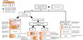 CMS algorithm.jpg 3,994 × 1,973; 179 KB
- UNIT OF COMPETENCY Lumbar TFIS.pdf ; 110 KB
 Reading man.jpg 2,048 × 1,280; 130 KB
 Region selector.jpg 1,029 × 2,059; 99 KB


