Spinal Cord Anatomy
The spinal cord starts below the foramen magnum, and ends at the tip of the conus medullaris. It ends between the T12 to L2-3 disc level, typically around L1 or the L1-2 disc. It is contained within the thecal sac. The spinal cord has a tubular shape. On cross section it is elliptical in the cervical region, and round in the thoracic region. There are two enlargements at the areas of limb innervation - the cervical enlargement at C5-T1 for upper limb innervation, and a smaller lumbar enlargement at T9-L2 for lower limb and pelvis innervation. There are 31 spinal segments along the spinal cord. These are the 8 cervical, 12 thoracic, five lumbar, five sacral, and one coccygeal segments. The filum terminalis is a thin band of connective tissue that extends from the tip of the conus medullaris down to the first coccygeal segment through the caudal end of the thecal sac.[1]
Fissures and Sulci

A deep ventral median fissure and a shallower dorsal median sulcus and septum posteriorly divides the spinal cord into right and left halves that are almost completely separated. Dorsal nerve roots travel to the cord on the posterior surface of the cord through the dorsolateral sulci. Ventral nerve roots travel from the anterior surface of the cord through the ventrolateral sulci. The topography of the cord surface can be visualised by axial T2 weighted gradient echo MRI sequences and CT myelograms.[1]
Grey Matter
Unlike the brain where the grey matter is located peripherally, the spinal cord has its grey matter centrally and is H- or butterfly-shaped. On MRI the grey matter is hyperintense on T2, and hypointense on T1. The peripheral white matter columns are hyperintense to grey on T1. The grey matter has ventral and dorsal horns that run along the length of the cord. There are also small lateral horns from around T1-L1. The dorsal horns receive primary afferent fibres from the dorsal roots of the spinal nerves and are primarily involved in sensory processing. The ventral horns transmit motor neurons. The lateral horns have autonomic nuclei and interneurons. Connecting the two arms of the H around the central canal are the ventral and dorsal gray matter commissures that run horizontally. The central canal is located between the ventral one-third and dorsal two-thirds in the cervical and thoracic region, and is more central in the lumbar region. On MRI it is a thin with a non-enhancing fluid signal.[1]
White Matter
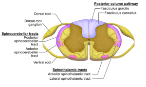
The white matter contains three columns otherwise known as funiculi. They are the ventral (anterior), lateral, and dorsal (posterior) columns. They contain nerve axon bundles making up the neural pathways. The ventral columns are located between the ventral median fissure and ventral nerve roots. The lateral columns are between the ventrolateral and dorsolateral sulci in the location of where the ventral nerve roots exit and the dorsal nerve roots enter the cord. The dorsal columns are between the dorsal median sulcus and dorsolateral sulci on each side. Through these columns travel the ascending sensory and descending motor pathways. The most well known columns are the corticospinal tract, the dorsal columns, and the spinothalamic tract.[1]
Spinal cord sensory pathways
Spinal cord motor pathways
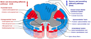
Spinal cord detailed pathways


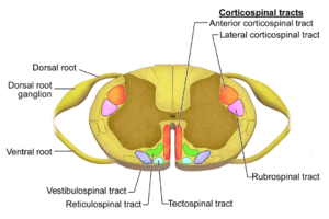
Corticospinal Tract

The corticospinal tract contains descending motor axons that control fine movements and the most important motor pathways. The axons are from upper motor neurons. More than 50% of these stem from the primary motor cortex, and the remainder come from the premotor cortex, supplementary motor area, and sensory cortex. From the brain they travel through the cerebral white matter, posterior limb of the internal capsule, cerebral peduncle, and the ventral pons. In the medulla, 75-90% of the nerve fibres decussate in the pyramid and then travel caudally becoming the lateral corticospinal tract in the lateral column. These lateral corticospinal tract neurons synapse in the ventral horn and then exit the spinal cord. The remaining axons that don't decussate, travel caudally as the anterior corticospinal tract. These fibres cross over to the contralateral side in each spinal cord segment, and supply motor neurons in the ventral horn.[1]
Injury to neurons in the motor cortex and corticospinal tract manifests as upper motor neuron deficits. While injury to the neurons in the ventral horns and peripheral nerves manifests as lower motor neuron deficits. There is a laminar somatotopic arrangement of axons that is similar in the corticospinal and spinothalamic tracts. Cervical and thoracic axons innervating the upper limbs and thorax are found medially. Lumbar and sacral axons innervating the abdomen and lower extremities are found laterally. This is why injury to the central spinal cord first affects the upper extremities.[1]
| Feature | UMN Deficit | LMN Deficit |
|---|---|---|
| Lesion location | Proximal to anterior horn (motor cortex, brainstem, spinal cord) | Anterior horn cell or distal to anterior horn cell (root, plexus, peripheral nerve) |
| Muscle tone | Increased (spastic paresis) | Decreased (flaccid paralysis) |
| Muscle bulk | Maintained or mildly atrophic | Severely atrophic |
| Weakness | In legs, greater in flexors than in extensors; in arms, greater in extensors than in flexors, producing a pyramidal pattern of weakness | Depends on location: uniform spinal cord weakness in all muscle groups of the segment; peripheral nerve weakness of specific muscle groups |
| Reflexes | Increased | Areflexia |
| Babinski sign | Positive: upgoing plantar response | Negative |
| Fasciculations | No | Yes |
Dorsal Columns
Spinothalamic Tract
Spinal Cord Blood Supply
References
Literature Review
 Literature Review
- Reviews from the last 7 years: review articles, free review articles, systematic reviews, meta-analyses, NCBI Bookshelf
- Articles from all years: PubMed search, Google Scholar search.
- TRIP Database: clinical publications about evidence-based medicine.
- Other Wikis: Radiopaedia, Wikipedia Search, Wikipedia I Feel Lucky, Orthobullets,

