Tarlov Cysts
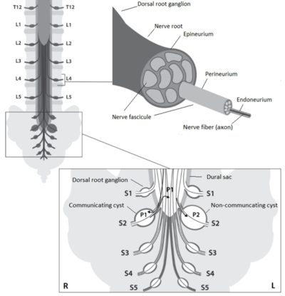
Tarlov cysts also called perineurial or sacral nerve root cysts are are dilations of the nerve root sheaths at the dorsal root ganglion. They manifest as abnormal sacs filled with cerebrospinal fluid. They are extradural but contain neural tissue.
It is a controversial topic whether they are a rare source of pain or often produce symptoms. The nonbelievers outweigh the believers.[1]
Differentiation
Spinal meningeal cysts are diverticula of the spinal meningeal sac, nerve root sheath, or arachnoid. Tarlov cysts are the most common type of meningeal cysts. They lie extradurally, near the dorsal root ganglion, and contain nerve fibres.
| Tarlov Cyst | Meningeal Diverticula & Arachnoid Diverticula |
|---|---|
| Potential communication with spinal subarachnoid space | Communicates freely with spinal subarachnoid space |
| Found distal to the junction of posterior nerve root and dorsal root ganglion in sacral region | Found proximal to dorsal root ganglion throughout vertebral column |
| Walls contain nerve fibers | Walls lined by arachnoid mater with no signs of neural element |
| Often multiple, extending around the circumference of nerve root | No pattern of formation in regards to multiplicity |
Pathogenesis and Pathophysiology

They are thought to arise through increased hydrostatic and pulsatile pressures from the spinal canal that force CSF into the nerve root sheaths. The cause of increased hydrostatic pressure isn't known. Tarlov cysts can be communicating (non-valved) or valved.
Initially the cyst is communicating, i.e. there is a connection with the subarachnoid space of the spinal canal, the so called cyst neck. The nerve fibres of the dorsal root ganglion are int he neck and spread over the cyst body or through the cavity.
Is some cases the neck can narrow and a one-way valve system is created which allows CSF to enter the cyst but limits its exit. Here the pressure rises to a point higher inside the cyst than in the spinal canal, and axons can be more compressed than in communicating Tarlov cysts. Valved cysts are usually larger than non-valved cysts.
Pulsatile and hydrodynamic forces of CSF cause these cysts to fill and expand in size. They occur most frequently in the sacrum where the hydrostatic pressure is the highest. Pain is thought to arise through increased hydrostatic pressure causing internal pressure which irritates and compresses the axons inside the nerve roots.[1]
In some patients Tarlov cysts are unnoticed as they become progressively larger and work as a buffer for excess hydrostatic pressure within the subarachnoid space. However when this buffer system decompensates or when a valve system is established it is thought that at this point patients may become symptomatic or more symptomatic.[1]
Aetiology
They can be acquired or congenital.
- It can occur due to trauma causing haemorrhage into the subarachnoid space. The accumulation of red cells may impede the drainage of the veins in the perineurium and epineurium, leading to rupture with subsequent cyst formation.
- Congenitally it can be caused by arachnoidal proliferations within the root sleeve.
One study found small and/or large fibre neuropathy in a significant proportion of patients with Tarlov cysts.[2]
Epidemiology
They are seen in up to 4.6% of the population, and are more common in women.[3] The prevalence was 13.2% in a cohort of patients who had MRI done for any reason.[4]
They are more commonly seen in patients with heritable connective tissue disorders, [1] including Marfan syndrome, Ehlers Danlos Syndrome, Sjogren syndrome, and Loeys-Dietz syndrome.
They are more prevalent in patients with perineal pain and persistent genital arousal disorder.[1]
Clinical Features
Most Tarlov cysts are thought to be asymptomatic. It is a controversial matter regarding the prevalence of symptomatic cases and what type of symptoms may occur. In some causes particularly with large cysts, neurological dysfunction and neuropathic pain can occur. However symptoms don't correlate very well with the size of the cyst.[5]
Possible symptoms include pelvic or perineal pain (S2-S4), sphincter or sexual dysfunction, radicular pain or radiculopathy, and neurogenic claudication.[6][7] Motor symptoms are less common because they primarily affect the dorsal root.[1]
When changing from lying to sitting, CSF volume is shifted caudally. In patients with non-valved cysts pain increases when sitting or standing, and is relieved when lying down. However in patients with valved cysts there is no relief with lying down due to limitation of CSF outflow.
S2, S3, S4
Sensory: perineum, clitoris, penis, vagina, scrotum
Autonomic: detrusor muscle of the bladder, descending colon, transverse colon, rectum, internal urinary sphincter, internal anal sphincter
Motor: external urinary sphincter, external anal sphincter
Symptoms: The patient may have pelvic and perineal pain with paraesthesias. Vaginal pain can occur with dyspareunia. In men testicular, penile, or prostate pain can occur. Neurogenic bladder and bowel symptoms may be seen, as well as erectile dysfunction and female anorgasmia.
S2
Sensory: perineum, posteromedial side of the legs, plantar region of the feet
Motor: intrinsic foot muscles
Symptoms: There may be atrophy of the intrinsic foot muscles due to compression of nerve root S2.
L5 and S1
Sensory: posterior side of the legs (S1), lateral side of the leg, first and fifth toe (L5), dorsal side of the feet
Motor: gluteus maximus muscle and calf muscles (S1), gluteus medius muscle and extensor muscles of the feet and toes (L5)
Symptoms: There may be lumbar, sacral, buttock, and lateral hip pain along with pain and/or paraesthesias in the legs and feet. Leg weakness can occur along with neurogenic claudication. There may be weakness of dorsiflexion of the foot and rarely foot drop for L5, and weakness of plantar flexion for S1.
L1 to L4
Paraesthesias and pain in the legs, weakness of the knee or hip extension
| Tarlov cysts | Valved cysts | Non-valved cysts |
|---|---|---|
| Size | Commonly >10mm | Commonly smaller |
| Onset of symptoms | Long pain-free interval | Onset early in life |
| Mean age of onset of symptoms | +/- 5th decade | +/- 2nd to 3rd decade |
| Evolution | Rapid progression | Slow progression |
| Sensory symptoms (pain and paraesthesia/numbness) | Commonly | Commonly |
| Motor symptoms (muscle weakness) | Commonly | Occasionally |
| Cauda equina syndrome | Acute | Chronic |
| Prevalence | Rare | Common |
| Reported in the literature | Commonly | Rarely |
| Clinical appearance | Easily diagnosed | Commonly overlooked |
| Surgical intervention | May be recommended in cases of severe pain or motor dysfunction | Probably not effective |
Imaging
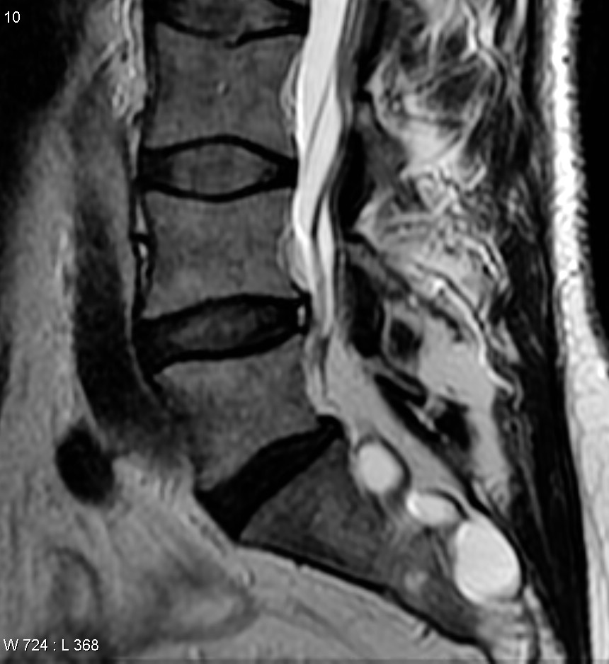
MRI is the imaging study of choice. They are most commonly seen in the lower lumbar spine and sacrum, however they can occur elsewhere in the spine.
They appear as very thin-walled CSF intensity (T1 low signal, T2 high signal) cystic structures that are closely related to the sacral and lower lumbar nerves. The sacral foramina may be widened. Morphology varies from a simple rounded cyst to a more complex loculated cystic mass.[8]
Differentials on imaging include dural ectasia, spinal synovial cyst, meningocele, nerve sheath tumour, and spinal metastases. [8]
On plain films there may be bony erosion of the spinal canal or of the sacral foramina
Diagnosis
Hulens and colleagues propose considering the following when determining if the cyst is symptomatic[1]
- Consider in patients with cysts on MRI with low back pain or radicular pain with no other obvious cause
- Sensory abnormalities, weakness, or hyporeflexia consistent with the location of the cyst
- Urodynamic testing showing early sensation of filling, detrusor contractions, urethral instability, stress incontinence.
- Electrophysiology (particularly L5 and sacral) showing delayed F waves, delayed S1 H-reflexes, delayed ano-anal reflexes, and neurogenic abnormalities during EMG
- Improved symptoms with lumbar puncture
Treatment
There is no consensus on the optimal treatment of symptomatic sacral perineural cysts. The following categories of treatment are all controversial.
Medication
Using acetazolamide to reduce CSF pressure and therefore reduce the hydrostatic pressure in the cyst has been used.[1]
Aspiration
Aspiration can be done under CT guidance using a two-needle technique. Fibrin can be injected to try seal the cyst. Complications include cyst recurrence, CSF leak, infection, and persistent procedural pain.
Lumbar Puncture
To reduce hydrostatic pressure.[1]
Surgery
There are various surgical approaches including microfenestration and surgical sleeving of the cysts, +/- biosynthetic dural patch, +/- muscle patch. Cyst excision results in irreversible neural damage.
References
- ↑ 1.00 1.01 1.02 1.03 1.04 1.05 1.06 1.07 1.08 1.09 1.10 Hulens M, Rasschaert R, Bruyninckx F, Dankaerts W, Stalmans I, De Mulder P, Vansant G. Symptomatic Tarlov cysts are often overlooked: ten reasons why-a narrative review. Eur Spine J. 2019 Oct;28(10):2237-2248. doi: 10.1007/s00586-019-05996-1. Epub 2019 May 11. PMID: 31079249.
- ↑ Hulens M, Bruyninckx F, Thal DR, Rasschaert R, Bervoets C, Dankaerts W. Large- and Small-Fiber Neuropathy in Patients with Tarlov Cysts. J Pain Res. 2022 Jan 25;15:193-202. doi: 10.2147/JPR.S342759. PMID: 35115823; PMCID: PMC8801331.
- ↑ Andrieux C, Poglia P, Laudato P. Tarlov Cyst: A diagnostic of exclusion. Int J Surg Case Rep. 2017;39:25-28. doi: 10.1016/j.ijscr.2017.07.045. Epub 2017 Jul 25. PMID: 28787671; PMCID: PMC5545870.
- ↑ Kuhn FP, Hammoud S, Lefèvre-Colau MM, Poiraudeau S, Feydy A. Prevalence of simple and complex sacral perineural Tarlov cysts in a French cohort of adults and children. J Neuroradiol. 2017 Feb;44(1):38-43. doi: 10.1016/j.neurad.2016.09.006. Epub 2016 Nov 9. PMID: 27836653.
- ↑ Davis SW, Levy LM, LeBihan DJ, Rajan S, Schellinger D. Sacral meningeal cysts: evaluation with MR imaging. Radiology. 1993 May;187(2):445-8. doi: 10.1148/radiology.187.2.8475288. PMID: 8475288.
- ↑ Marino D, Carluccio MA, Di Donato I, Sicurelli F, Chini E, Di Toro Mammarella L, Rossi F, Rubegni A, Federico A. Tarlov cysts: clinical evaluation of an italian cohort of patients. Neurol Sci. 2013 Sep;34(9):1679-82. doi: 10.1007/s10072-013-1321-0. Epub 2013 Feb 12. PMID: 23400656.
- ↑ Hulens Mieke, Bruyninckx Frans, Somers Alix, Stalmans Ingeborg, Peersman Benjamin, Vansant Greet, Ricky Rasschaert, De Mulder Peter, Dankaerts Wim. Electromyography and A Review of the Literature Provide Insights into the Role of Sacral Perineural Cysts in Unexplained Chronic Pelvic, Perineal and Leg Pain Syndromes. (2017) International Journal of Physical Medicine & Rehabilitation. 5 (3): 1. doi:10.4172/2329-9096.1000407
- ↑ 8.0 8.1 Jones, J., Bell, D. Tarlov cyst. Reference article, Radiopaedia.org. (accessed on 14 Feb 2022) https://doi.org/10.53347/rID-10198
Literature Review
- Reviews from the last 7 years: review articles, free review articles, systematic reviews, meta-analyses, NCBI Bookshelf
- Articles from all years: PubMed search, Google Scholar search.
- TRIP Database: clinical publications about evidence-based medicine.
- Other Wikis: Radiopaedia, Wikipedia Search, Wikipedia I Feel Lucky, Orthobullets,


