File:Finger extensor mechanism.jpg
Finger_extensor_mechanism.jpg (610 × 457 pixels, file size: 64 KB, MIME type: image/jpeg)
Summary
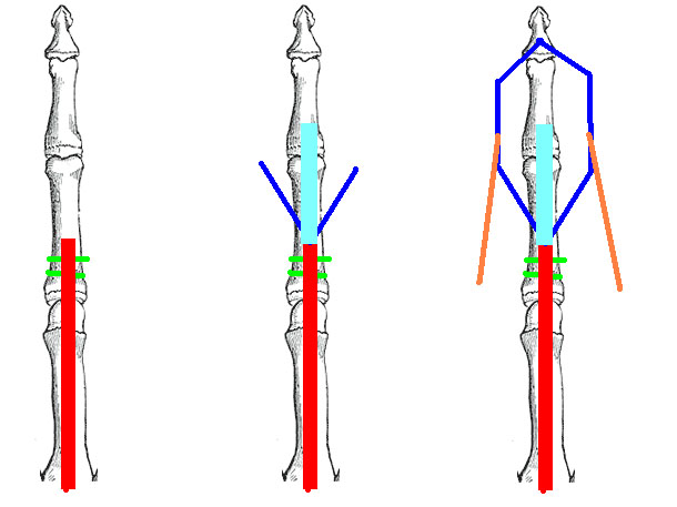
Extensor mechanism of the finger. In the drawing on the left the common extensor tendon is shown in red. It attaches to the proximal phalanx via bands and extends the MCP joint. In the center figure, the trifurcation is shown. the central slip, shown in sky blue, inserts on the middle phalanx and extends the PIP joint. The lateral bands are shown in blue. In the right-most drawing, the lateral bands are shown joined by the tendons of the interosseous and lumbrical muscles and inserting on the distal phalanx, thereby extending the DIP.
From https://orthopaedia.com/page/Mallet-finger-and-other-finger-extensor-injuries
Licencing
![]()
This work is licensed under the Creative Commons Attribution-NonCommercial-ShareAlike License.
File history
Click on a date/time to view the file as it appeared at that time.
| Date/Time | Thumbnail | Dimensions | User | Comment | |
|---|---|---|---|---|---|
| current | 20:51, 3 April 2022 | | 610 × 457 (64 KB) | Jeremy (talk | contribs) | Extensor mechanism of the finger. In the drawing on the left the common extensor tendon is shown in red. It attaches to the proximal phalanx via bands and extends the MCP joint. In the center figure, the trifurcation is shown. the central slip, shown in sky blue, inserts on the middle phalanx and extends the PIP joint. The lateral bands are shown in blue. In the right-most drawing, the lateral bands are shown joined by the tendons of the interosseous and lumbrical muscles and inserting on the... |
You cannot overwrite this file.
File usage
The following page uses this file:

