File:Greater trochanter anatomy facets insertions and bursae.jpg
Greater_trochanter_anatomy_facets_insertions_and_bursae.jpg (768 × 369 pixels, file size: 48 KB, MIME type: image/jpeg)
Summary
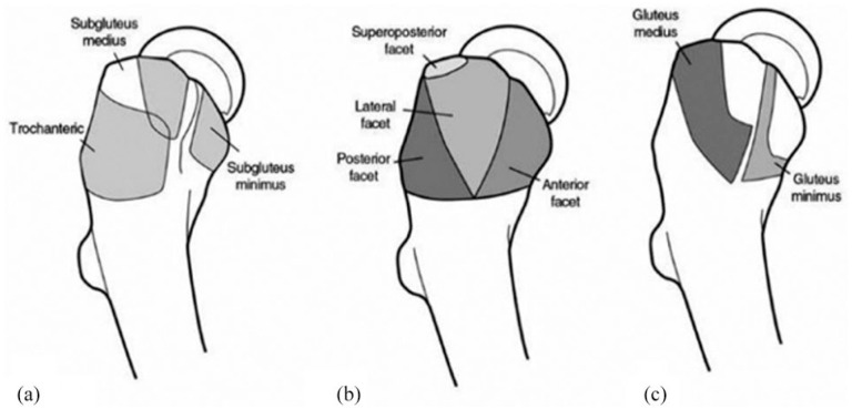
Anatomy of the greater trochanter. (a) Three peritrochanteric bursae, (b) osseous facets of the greater trochanter, and (c) insertion sites for the abductor tendons
From Pianka MA, Serino J, DeFroda SF, Bodendorfer BM. Greater trochanteric pain syndrome: Evaluation and management of a wide spectrum of pathology. SAGE Open Med. 2021 Jun 3;9:20503121211022582. doi: 10.1177/20503121211022582. PMID: 34158938; PMCID: PMC8182177.
Licensing
![]()
This work is licensed under the Creative Commons Attribution-NonCommercial 4.0 License.
File history
Click on a date/time to view the file as it appeared at that time.
| Date/Time | Thumbnail | Dimensions | User | Comment | |
|---|---|---|---|---|---|
| current | 19:52, 11 April 2022 | | 768 × 369 (48 KB) | Jeremy (talk | contribs) | Anatomy of the greater trochanter. (a) Three peritrochanteric bursae, (b) osseous facets of the greater trochanter, and (c) insertion sites for the abductor tendons From Pianka MA, Serino J, DeFroda SF, Bodendorfer BM. Greater trochanteric pain syndrome: Evaluation and management of a wide spectrum of pathology. SAGE Open Med. 2021 Jun 3;9:20503121211022582. doi: 10.1177/20503121211022582. PMID: 34158938; PMCID: PMC8182177. |
You cannot overwrite this file.
File usage
The following page uses this file:

