File:Acetabular version.png
From WikiMSK
No higher resolution available.
Acetabular_version.png (476 × 291 pixels, file size: 107 KB, MIME type: image/png)
Summary
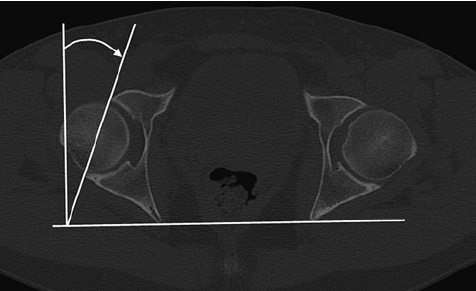
From Dandachli W, Ul Islam S, Tippett R, Hall-Craggs MA, Witt JD. Analysis of acetabular version in the native hip: comparison between 2D axial CT and 3D CT measurements. Skeletal Radiol. 2011 Jul;40(7):877-83. doi: 10.1007/s00256-010-1065-3. Epub 2010 Dec 22. PMID: 21181403.
Licencing
![]() This file is copyrighted, and is reproduced in a limited way under the fair-use doctrine. It falls under the "Non-profit educational" clause of the Fair Use doctrine.
This file is copyrighted, and is reproduced in a limited way under the fair-use doctrine. It falls under the "Non-profit educational" clause of the Fair Use doctrine.
File history
Click on a date/time to view the file as it appeared at that time.
| Date/Time | Thumbnail | Dimensions | User | Comment | |
|---|---|---|---|---|---|
| current | 18:24, 15 September 2021 | | 476 × 291 (107 KB) | Jeremy (talk | contribs) | From Dandachli W, Ul Islam S, Tippett R, Hall-Craggs MA, Witt JD. Analysis of acetabular version in the native hip: comparison between 2D axial CT and 3D CT measurements. Skeletal Radiol. 2011 Jul;40(7):877-83. doi: 10.1007/s00256-010-1065-3. Epub 2010 Dec 22. PMID: 21181403. |
You cannot overwrite this file.
File usage
The following page uses this file:

