File:Enthesis structure.png
Original file (850 × 871 pixels, file size: 240 KB, MIME type: image/png)
Summary
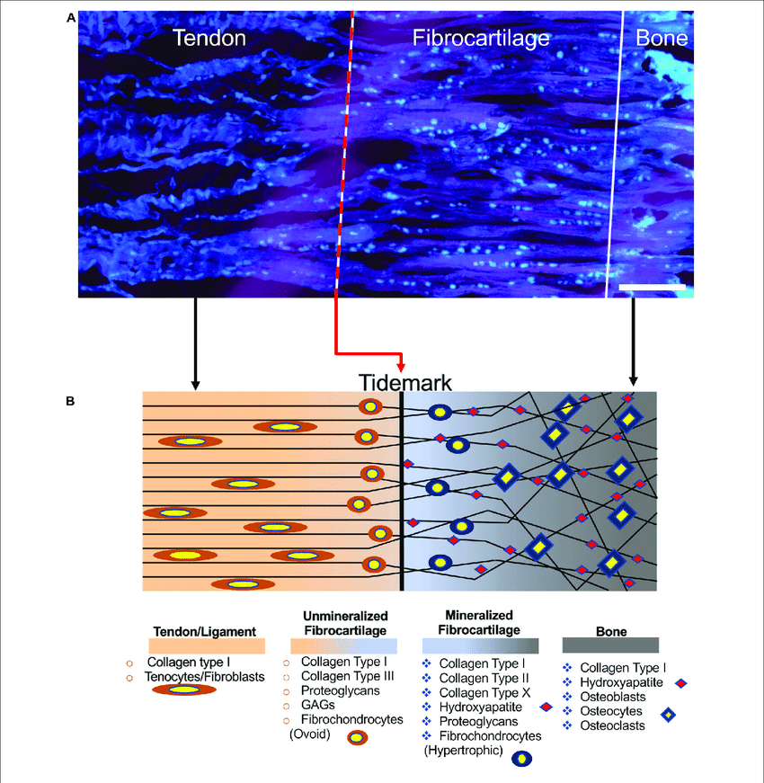
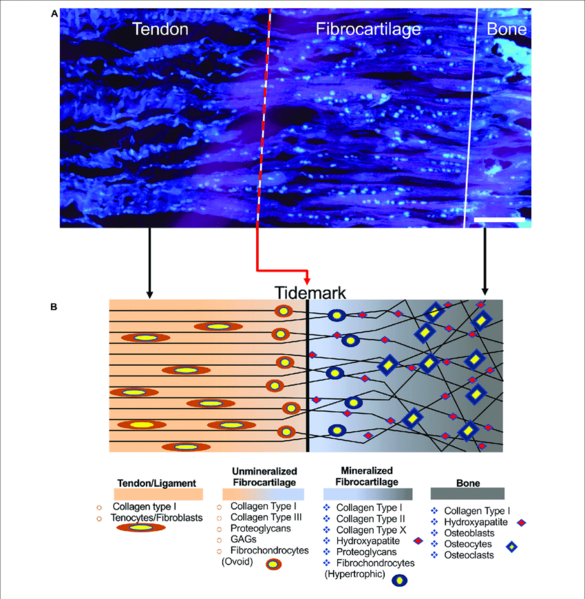
Structure of the enthesis. (A) Enthesis cryocut section of a porcine Achilles tendon was stained for cells using SYTO R 13. Cells are depicted cyan (scale bar = 150 µm). (B) Graphical representation of the enthesis and its components.
From Sensini, Alberto et al. “Tissue Engineering for the Insertions of Tendons and Ligaments: An Overview of Electrospun Biomaterials and Structures.” Frontiers in bioengineering and biotechnology vol. 9 645544. 2 Mar. 2021, doi:10.3389/fbioe.2021.645544
Licencing
![]()
This work is licensed under a Creative Commons Attribution 4.0 International License.
File history
Click on a date/time to view the file as it appeared at that time.
| Date/Time | Thumbnail | Dimensions | User | Comment | |
|---|---|---|---|---|---|
| current | 18:41, 9 August 2021 | | 850 × 871 (240 KB) | Jeremy (talk | contribs) | From Sensini, Alberto et al. “Tissue Engineering for the Insertions of Tendons and Ligaments: An Overview of Electrospun Biomaterials and Structures.” Frontiers in bioengineering and biotechnology vol. 9 645544. 2 Mar. 2021, doi:10.3389/fbioe.2021.645544 |
You cannot overwrite this file.
File usage
The following page uses this file:

