File:Sagittal band tear.jpg
From WikiMSK
Size of this preview: 800 × 431 pixels. Other resolution: 979 × 527 pixels.
Original file (979 × 527 pixels, file size: 54 KB, MIME type: image/jpeg)
Summary
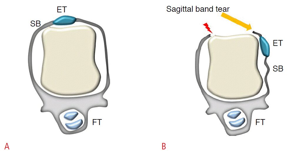
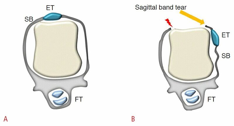
Schematic images of the sagittal band and sagittal band tears.
A, B. At the metacarpophalangeal joint, the extensor tendons are stabilized by the extensor hood and particularly by the sagittal band (A). Subluxation or dislocation of the extensor tendon occurs as a result of a tear in the sagittal band (B). ET, extensor tendon; FT, flexor tendon; SB, sagittal band.[1]
Licencing
![]()
This work is licensed under the Creative Commons Attribution-NonCommercial 4.0 License.
- ↑ Lee SA, Kim BH, Kim SJ, Kim JN, Park SY, Choi K. Current status of ultrasonography of the finger. Ultrasonography. 2016 Apr;35(2):110-23. doi: 10.14366/usg.15051. Epub 2015 Nov 24. PMID: 26753604; PMCID: PMC4825212.
File history
Click on a date/time to view the file as it appeared at that time.
| Date/Time | Thumbnail | Dimensions | User | Comment | |
|---|---|---|---|---|---|
| current | 12:24, 6 February 2022 | | 979 × 527 (54 KB) | Jeremy (talk | contribs) | Lee SA, Kim BH, Kim SJ, Kim JN, Park SY, Choi K. Current status of ultrasonography of the finger. Ultrasonography. 2016 Apr;35(2):110-23. doi: 10.14366/usg.15051. Epub 2015 Nov 24. PMID: 26753604; PMCID: PMC4825212. |
You cannot overwrite this file.
File usage
The following page uses this file:

