Hip Joint: Difference between revisions
| Line 23: | Line 23: | ||
The acetabulum is horseshoe shaped with the acetabular notch in the inferior region. The acetabular cartilage is thicker in the periphery where it merges with the labrum. It is particularly thick in the superior-anterior region of the dome. Articulation with the femoral head only occurs on the horseshoe-shaped hyaline cartilage. | The acetabulum is horseshoe shaped with the acetabular notch in the inferior region. The acetabular cartilage is thicker in the periphery where it merges with the labrum. It is particularly thick in the superior-anterior region of the dome. Articulation with the femoral head only occurs on the horseshoe-shaped hyaline cartilage. | ||
The acetabulum is angled obliquely forward: it is directed anterior, lateral, and inferior. A mal-aligned acetabulum doesn't adequately cover the femoral head and this can lead to chronic dislocation and early onset osteoarthritis. | |||
The acetabulum is angled obliquely forward: it is directed anterior, lateral, and inferior. A mal-aligned acetabulum doesn't adequately cover the femoral head and this can lead to chronic dislocation and early onset osteoarthritis. | |||
How much coverage the acetabulum provides over the femoral head in the coronal plane is measured by various parameters on radiographs such as the centre edge angle which normally measures 20-45° (Figure 1). A normal centre edge angle provides a protective shelf over the femoral head in the coronal plane, while a small angle means that there is less coverage leading to risk of dislocation. | How much coverage the acetabulum provides over the femoral head in the coronal plane is measured by various parameters on radiographs such as the centre edge angle which normally measures 20-45° (Figure 1). A normal centre edge angle provides a protective shelf over the femoral head in the coronal plane, while a small angle means that there is less coverage leading to risk of dislocation. | ||
In the horizontal plane coverage can be measured by angle of acetabular anteversion and measures 20° on average. An increase in this angle is associated with decreased joint stability and an increased risk of anterior dislocation.(See [[Hip Radiograph]]) | In the horizontal plane coverage can be measured by angle of acetabular anteversion and measures 20° on average (Figure 2). An increase in this angle is associated with decreased joint stability and an increased risk of anterior dislocation.(See [[Hip Radiograph]]) | ||
<gallery mode=packed heights=300px> | <gallery mode=packed heights=300px> | ||
Acetabular lateral centre edge and acetabular index.jpg|'''Figure 1'''. The lateral centre-edge angle (LCEA) and acetabular index (AI) on AP pelvic radiograph are measures of coverage of the femoral head by the acetabulum. | Acetabular lateral centre edge and acetabular index.jpg|'''Figure 1'''. The lateral centre-edge angle (LCEA) and acetabular index (AI) on AP pelvic radiograph are measures of coverage of the femoral head by the acetabulum. | ||
Acetabular version.png|'''Figure 2''' The acetabular version angle can be measured on CT in the axial plane. The acetabulum is normally anteverted to around 20°. | Acetabular version.png|'''Figure 2''' The acetabular version angle can be measured on CT in the axial plane. The acetabulum is normally anteverted to around 20°. | ||
</gallery> | </gallery>The labrum is a fibrocartilaginous lip that encircles and deepens the acetabulum. It blends with the transverse acetabular ligament which spans the acetabular notch. It helps to contain the femoral head in place in extremes of motion, especially in flexion. Along with the capsule, the labrum acts as a load-bearing structure during flexion. The labrum helps to maintain a negative pressure vacuum within the joint that is produced by the acetabular fossa which helps to stabilise the joint. Individuals with a [[Hip Labral Tear|deficient labrum]] may have instability and capsular laxity. | ||
=== Femoral Head === | === Femoral Head === | ||
Revision as of 18:31, 15 September 2021
{{{{{quality}}}}}
| Hip Joint | |
|---|---|
| Primary Type | Ball and socket"Ball and socket" is not in the list (Synovial Joint, Cartilaginous Joint, Fibrous Joint, Compound Joint) of allowed values for the "Has joint type" property. |
| Secondary Type | |
| Bones | Ilium, femur |
| Ligaments | Iliofemoral, ischiofemoral, ligamentum teres |
| Muscles | |
| Innervation | |
| Vasculature | |
| ROM | |
| Volume | |
| Conditions | |
Bones and Articulations
The hip is a ball and socket joint. The ball is the spherical head of the femur, while the socket is the concave acetabulum.
Acetabulum
The acetabulum is horseshoe shaped with the acetabular notch in the inferior region. The acetabular cartilage is thicker in the periphery where it merges with the labrum. It is particularly thick in the superior-anterior region of the dome. Articulation with the femoral head only occurs on the horseshoe-shaped hyaline cartilage.
The acetabulum is angled obliquely forward: it is directed anterior, lateral, and inferior. A mal-aligned acetabulum doesn't adequately cover the femoral head and this can lead to chronic dislocation and early onset osteoarthritis.
How much coverage the acetabulum provides over the femoral head in the coronal plane is measured by various parameters on radiographs such as the centre edge angle which normally measures 20-45° (Figure 1). A normal centre edge angle provides a protective shelf over the femoral head in the coronal plane, while a small angle means that there is less coverage leading to risk of dislocation.
In the horizontal plane coverage can be measured by angle of acetabular anteversion and measures 20° on average (Figure 2). An increase in this angle is associated with decreased joint stability and an increased risk of anterior dislocation.(See Hip Radiograph)
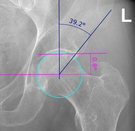
Figure 1. The lateral centre-edge angle (LCEA) and acetabular index (AI) on AP pelvic radiograph are measures of coverage of the femoral head by the acetabulum.
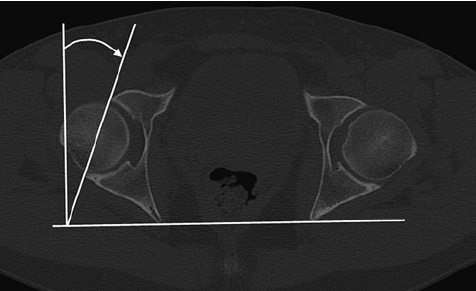
Figure 2 The acetabular version angle can be measured on CT in the axial plane. The acetabulum is normally anteverted to around 20°.


The labrum is a fibrocartilaginous lip that encircles and deepens the acetabulum. It blends with the transverse acetabular ligament which spans the acetabular notch. It helps to contain the femoral head in place in extremes of motion, especially in flexion. Along with the capsule, the labrum acts as a load-bearing structure during flexion. The labrum helps to maintain a negative pressure vacuum within the joint that is produced by the acetabular fossa which helps to stabilise the joint. Individuals with a deficient labrum may have instability and capsular laxity.
Femoral Head
The femur is the largest and strongest bone in the body. The femoral neck is the narrowest part and internally it is primarily composed of trabecular bone, and so it is the weakest component of the femur. The femur angles medially downward to aid in single-leg support during walking and running.
Stability
The labrum is a ring of fibrocartilage that contributes to joint stability. Compared to the glenoid fossa of the shoulder, the acetabular fossa is much deeper and so is more stable.
There are several strong ligaments that contribute to stability.
- Anterior ligaments: Iliofemoral or Y ligament plus the pubofemoral ligament
- Posterior ligaments: ischiofemoral ligament
- Inferior ligaments: Transverse acetabular ligament, prevents inferior dislocation of the femoral head.
These major ligaments act to twist the head of the femur into the acetabulum during hip extension.
Within the joint capsule is the ligamentum teres which attaches directly from the rim of the acetabulum to the head of the femur.
Bursae
The iliopsoas bursa and deep trochanteric bursa are the most prominent bursae. The iliopsoas is located between the iliopsoas and articular capsule and reduces friction between these two structures. The deep trochanteric bursa reduces friction between the greater trochanter of the femur and the gluteus maximus.
Movements
The pelvic girdle is analogous to the shoulder girdle in that it positions the hip for effective movement. The shoulder girdle is made up of multiple different joints, while the pelvis is non-jointed. Nevertheless, the pelvis is able to rotate in all three planes. With pelvic rotation, the acetabular position is changed so that it is directed towards the impending femoral movement. Posterior pelvic tilt enables increases hip flexion by placing the head of the femur in front of the pelvis. Anterior pelvic tilt increases hip extension, and lateral pelvic tilt increases lateral movements. The pelvis also moves in coordination with the spine.
There are a variety of two-joint muscles. Two-joint muscles function most effectively at one joint when the position of the other joint stretches the muscle slightly.
- Flexion: iliacus, psoas major, pectineus, rectus femoris, sartorius, and tensor fascia lata.
- Iliopsoas (iliacus and psoas major) is the major hip flexor.
- Rectus femoris is a two-joint muscle and is active during both hip flexion and knee extension; it works better as a hip flexor with the knee is flexion.
- Sartorius is also a two-joint muscle.
- Extension: Gluteus maximus, biceps femoris, semitendinosus, and semimembranosus (latter three are the hamstrings).
- Gluteus maximus is active only with hip flexion
- Hamstrings are two-joint muscles, and aid in hip extension and knee flexion.
- Abduction: Gluteus medius and minimus
- Gluteus medius: major abductor
- Gluteus minimus: assists.
- Both muscles stabilise the pelvis during the support phase of walking and running and with standing on one leg.
- Adduction:Adductor longus, adductor brevis, adductor magnus, gracilis
- Active during swing phase of gait to bring the foot below the body's centre of gravity before the support phase. Also active during stair and hill climbing.
- Gracilis: strap muscle contributes to knee flexion.
- Adductor longus, adductor brevis, adductor magnus: also contributes to flexion and lateral hip rotation especially with a medially rotated femur.
- Lateral rotators: piriformis, gemellus superior, gemellus inferior, obturator internus, obturator externus, quadratus femoris
- Stronger than medial rotators.
- Medial rotators: gluteus minimus, tensor fascia lata, semitendinosus, semimembranosus, gluteus medius
- Horizontal abduction
- Horizontal abduction

