Hip Radiograph: Difference between revisions
No edit summary |
|||
| (5 intermediate revisions by the same user not shown) | |||
| Line 29: | Line 29: | ||
*[[Pelvic Radiograph|Pelvic AP radiograph]] | *[[Pelvic Radiograph|Pelvic AP radiograph]] | ||
*Dunn view | *Dunn view | ||
** | **lateral projection to aid and diagnose [[Femoroacetabular Impingement|femoroacetabular impingement]] (FAI) due to its increased sensitivity for detecting femoral head-neck asphericity. | ||
*frog leg view | *frog leg view | ||
**bilateral examination allows for better visualisation of the hip joints and femoral neck. It is almost exclusively used in the paediatric population to assess for slipped upper femoral epiphysis (SUFE) and Perthes disease. | **bilateral examination allows for better visualisation of the hip joints and femoral neck. It is almost exclusively used in the paediatric population to assess for slipped upper femoral epiphysis (SUFE) and Perthes disease. | ||
==Interpretation== | ==Interpretation== | ||
Many of the measurements can be conceptualised as follows in the frontal and horizontal plane. | |||
* Acetabulum coverage frontal plane - lateral centre edge etc | |||
* Acetabulum coverage horizontal plane - acetabular version. However there are some signs on the frontal plane (crossover sign etc). | |||
* Femoral neck frontal plane - angle of inclination | |||
* Femoral neck horizontal plane - [[:File:Femoral neck torsion.png|femoral neck torsion]]. | |||
Acetabular morphology is assessed by looking at the following | |||
*Orientation – Posterior wall sign, crossover sign, ischial spine sign | |||
*Depth – coxa profunda, protrusion acetabuli, acetabular depth | |||
*Coverage – L-CEA, A-CEA, tonnis angle, sharp angle, extrusion index | |||
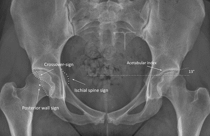
===Acetabular Orientation and Morphology=== | |||
[[File:Hip AP view radiographic signs.png|600px]] | [[File:Hip AP view radiographic signs.png|600px]] | ||
<gallery mode=packed heights=250px> | |||
File:Coxa profunda xray.jpg|Coxa profunda | |||
File:protrusio acetabuli xray.jpg|Protrusio acetabuli | |||
File:crossover posterior wall and ischial spine signs xray.png|Crossover sign, posterior wall sign, and ischial spine signs | |||
</gallery> | |||
===Acetabular Depth and Coverage Calculations=== | |||
Normal is 75% coverage | |||
#Acetabular depth ratio | |||
#Lateral centre edge angle L-CEA – normal 20-45 (Wiberg variant W-CEA is to edge of sourcil, 18-42). Confusingly the W-CEA is connected with Ogata et al, and the L-CEA with wiberg | |||
#Femoral extrusion – normal less than 25% | |||
#Lequesnes anterior centre edge angle – normal 25-50 | |||
<gallery mode=packed heights=250px> | |||
File:Acetabular coverage depth AP film.png|Acetabular depth ratio (1), lateral centre edge angle (2), femoral extrusion (3) | |||
File:Lequesnes anterior centre edge angle ap film.jpg|Lequenses anterior centre edge angle | |||
</gallery> | |||
#Tonnis Angle – draw towards most medial sclerotic edge (the sourcil). | |||
#*Normal: 0-10 degrees | |||
#*Dysplasia: >10 degrees | |||
#*Pincer FAI risk: <0 degrees | |||
#Sharp angle – draw from distal part of teardrop (total acetabulum) | |||
#*Dysplasia: >45 degrees | |||
<gallery mode=packed heights=250px> | |||
File:Acetabular coverage angle AP film.png|Tonnis angle (1), sharp angle (2) | |||
</gallery> | |||
== Further Reading == | |||
* Lim et al open access review article<ref>Lim SJ, Park YS. Plain Radiography of the Hip: A Review of Radiographic Techniques and Image Features. Hip Pelvis. 2015 Sep;27(3):125-34. doi: 10.5371/hp.2015.27.3.125. Epub 2015 Sep 30. PMID: 27536615; PMCID: PMC4972716.</ref> | |||
* Sutter et al open access review article.<ref>{{#pmid:22919039}}</ref> | |||
==References== | ==References== | ||
| Line 47: | Line 92: | ||
[[Category:Hip Joint]] | [[Category:Hip Joint]] | ||
[[Category:Radiology]] | [[Category:Radiology]] | ||
Latest revision as of 20:27, 15 April 2022

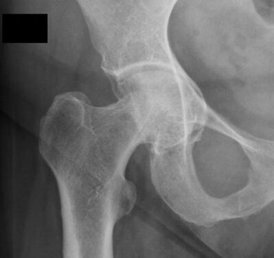
The hip series is comprised of an anteroposterior (AP) and lateral radiograph of the hip joint. The series is requested for a myriad of reasons from trauma to atraumatic hip pain.
Indications
Hip radiographs are performed for a variety of indications including
- trauma
- hip pain
- abnormal gait
- inability to weight bear
- arthropathy
- knee pain
Projections
Standard projections
- AP view
- demonstrates the hip joint in the AP plane, with the limb internally rotated so the neck of the femur is in profile
- often only performed in follow up studies
- lateral view
- standard rolled lateral view demonstrating the femoral neck and acetabular rim can only be performed on non-trauma patients
Modified trauma projections
- horizontal beam lateral view
- lateral projection demonstrating the neck of the femur without movement of the affected limb
- can only be conducted on unilateral hip trauma
- Clements-Nakayama view
- lateral projection demonstrating the neck of the femur without movement of the either limb
- the ideal projection for bilateral hip or femur trauma
Additional projections
- Pelvic AP radiograph
- Dunn view
- lateral projection to aid and diagnose femoroacetabular impingement (FAI) due to its increased sensitivity for detecting femoral head-neck asphericity.
- frog leg view
- bilateral examination allows for better visualisation of the hip joints and femoral neck. It is almost exclusively used in the paediatric population to assess for slipped upper femoral epiphysis (SUFE) and Perthes disease.
Interpretation
Many of the measurements can be conceptualised as follows in the frontal and horizontal plane.
- Acetabulum coverage frontal plane - lateral centre edge etc
- Acetabulum coverage horizontal plane - acetabular version. However there are some signs on the frontal plane (crossover sign etc).
- Femoral neck frontal plane - angle of inclination
- Femoral neck horizontal plane - femoral neck torsion.
Acetabular morphology is assessed by looking at the following
- Orientation – Posterior wall sign, crossover sign, ischial spine sign
- Depth – coxa profunda, protrusion acetabuli, acetabular depth
- Coverage – L-CEA, A-CEA, tonnis angle, sharp angle, extrusion index
Acetabular Orientation and Morphology

Coxa profunda

Protrusio acetabuli
Crossover sign, posterior wall sign, and ischial spine signs


Acetabular Depth and Coverage Calculations
Normal is 75% coverage
- Acetabular depth ratio
- Lateral centre edge angle L-CEA – normal 20-45 (Wiberg variant W-CEA is to edge of sourcil, 18-42). Confusingly the W-CEA is connected with Ogata et al, and the L-CEA with wiberg
- Femoral extrusion – normal less than 25%
- Lequesnes anterior centre edge angle – normal 25-50
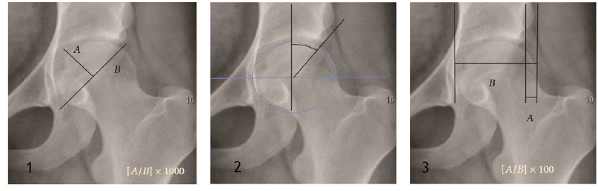
Acetabular depth ratio (1), lateral centre edge angle (2), femoral extrusion (3)
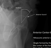
Lequenses anterior centre edge angle


- Tonnis Angle – draw towards most medial sclerotic edge (the sourcil).
- Normal: 0-10 degrees
- Dysplasia: >10 degrees
- Pincer FAI risk: <0 degrees
- Sharp angle – draw from distal part of teardrop (total acetabulum)
- Dysplasia: >45 degrees
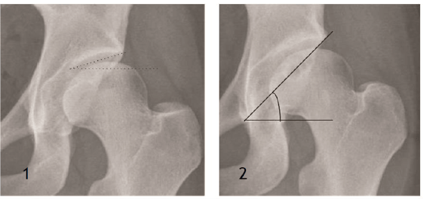
Tonnis angle (1), sharp angle (2)

Further Reading
References
Part or all of this article or section is derived from Hip series by Amanda Er and Andrew Murphy et al., used under CC BY-NC-SA 3.0
- ↑ Lim SJ, Park YS. Plain Radiography of the Hip: A Review of Radiographic Techniques and Image Features. Hip Pelvis. 2015 Sep;27(3):125-34. doi: 10.5371/hp.2015.27.3.125. Epub 2015 Sep 30. PMID: 27536615; PMCID: PMC4972716.
- ↑ Sutter et al.. New developments in hip imaging. Radiology 2012. 264:651-67. PMID: 22919039. DOI.

