Ligaments of the Foot and Ankle

The ligaments that surround the ankle act to limit plantarflexion and dorsiflexion, anterior and posterior movement of the foot, tilting of the talus, and inversion and eversion. Each of the different lateral ligaments have different roles in ankle stabilisation that depends on the position of the foot. Ankle stability depends on ligament orientation, loading type, and ankle position at the point of stress. The lateral ankle is more susceptible to injury.[1]
Overview
| Ligament | Insertion | Action |
|---|---|---|
| Anterior talofibular | Lateral malleolus TO neck of talus | Limits anterior displacement of foot or talar |
| Anterior talotibial | Anterior margin of tibia TO front margin on talus | Limits plantarflexion and abduction of foot |
| Calcaneocuboid | Calcaneus TO cuboid on dorsal surface | Limits inversion of foot |
| Calcaneofibular | Lateral malleolus TO tubercle on outer calcaneus | Resists backward displacement of foot; resists inversion |
| Deltoid | Medial malleolus TO talus, navicular, calcaneus | Resists valgus forces to ankle; limits plantarflexion, dorsiflexion, eversion, abduction of foot |
| Dorsal (tarsometatarsal) | Tarsals TO metatarsals | Supports arch; maintains relationship between tarsals and metatarsal |
| Dorsal calcaneocuboid | Calcaneus TO cuboid on dorsal side | Limits inversion |
| Dorsal talonavicular | Neck of talus TO superior surface of navicular | Supports talonavicular joint; limits inversion |
| Interosseous (intertarsal) | Connects adjacent tarsals | Supports arch of foot, intertarsal joints |
| Interosseous (talocalcaneal) | Undersurface of talus TO upper surface of calcaneus | Limits pronation, supination, abduction, adduction, dorsiflexion, plantarflexion |
| Plantar calcaneocuboid | Undersurface of calcaneus TO undersurface of cuboid | Supports arch |
| Plantar calcaneonavicular (spring) | Anterior margin of calcaneus TO undersurface on navicular | Supports arch; limits abduction |
| Posterior talofibular | Inner, back lateral malleolus TO posterior surface of talus | Limits plantarflexion, dorsiflexion, inversion; supports lateral ankle |
| Posterior talotibial | Tibial TO talus behind articulating facet | Limits planatarflexion; supports medial ankle |
| Talocalcaneal | Connecting anterior/posterior, medial, lateral talus TO calcaneus | Supports subtalar joint |
Deltoid Ligament
The complex of the medial collateral ligaments of the ankle joint is collectively called deltoid ligament. It attaches the medial malleolus to multiple tarsal bones.
The ligament is composed of two layers. The superficial layer has variable attachments and crosses two joints while the deep layer has talar attachments and crosses one joint:
It is a strong triangular band attached to the apex and the anterior and posterior borders of the medial malleolus. The plantar calcaneonavicular ligament can be considered as part of the medial ligament complex.
- superficial layer (from anterior to posterior)
- tibionavicular ligament (anterior)
- pass forwards to the navicular tuberosity behind which they blend with the medial margin of the plantar calcaneonavicular ligament
- tibiospring ligament
- descends vertically and blends with the fibres of the spring ligament
- tibiocalcaneal ligament (intermediate)
- descend almost vertically to the entire length of the sustentaculum tali
- superficial posterior tibiotalar ligament (posterior)
- pass posterolateral to the medial side of the talus and its medial tubercle
- tibionavicular ligament (anterior)
- deep layer: this layer is intra-articular and is covered by synovium
- anterior tibiotalar ligament (ATTL)
- short and thin ligament, pass from the tip of the medial malleolus to the non-articular part of the medial talar surface
- crossed by the tendons of the tibialis posterior and flexor digitorum longus
- deep posterior tibiotalar ligament (DPTTL)
- large and strong ligament from the medial malleolus to the talus
- covered by the superficial posterior tibiotalar and tibiocalcaneal ligaments
- anterior tibiotalar ligament (ATTL)
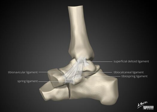
Deltoid superficial layer
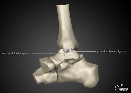
Deltoid deep layer


Part or all of this article or section is derived from Deltoid ligament of the ankle by Assoc Prof Craig Hacking and Dr Yuranga Weerakkody et al., used under CC BY-NC-SA 3.0
Lateral Collateral Ligament

The lateral collateral ligament (complex) of the ankle is a set of three ligaments that resist inversion of the ankle joint. They are more commonly injured than the medial collateral (deltoid) ligament of the ankle. They run from the lateral malleolus of the fibula to the talus and calcaneus.
The fan-like arrangement is composed of three ligaments:
- anterior talofibular ligament (ATFL)
- anterior component
- extends anteromedially from the anterior margin of the fibular malleolus to the talus, attached inferior of its lateral articular facet and to the lateral aspect of its neck
- under greatest tension in plantarflexion
- resists ankle inversion in plantarflexion, resistance to anterior talar displacement from the mortise (anterior drawer), and resistance to internal rotation of the talus within the mortise.
- calcaneofibular ligament
- middle component
- runs from the depression anterior to the apex of the fibular malleolus to a tubercle on the lateral calcaneal surface
- crossed by fibularis longus and brevis
- under greatest tension in dorsiflexion
- resists ankle inversion in dorsiflexion, also contributes to subtalar joint stability.
- posterior talofibular ligament (PTFL)
- posterior component
- runs horizontally from the distal part of the lateral malleolar fossa to the lateral tubercle of the posterior talar process
- a tibial slip of fibres connects it to the medial malleolus
- under greatest strain in dorsiflexion
- resists posterior talar displacement within the mortise, and limits talar external rotation.
- the syndesmosis can be considered as part of the lateral complex
- anterior inferior tibiofibular ligament (AITFL)
- posterior inferior tibiofibular ligament (PITFL)
- transverse tibiofibular ligament (TTFL)
- interosseous ligament
Part or all of this article or section is derived from Lateral collateral ligament of the ankle by Dr Yuranga Weerakkody and Dr Henry Knipe et al., used under CC BY-NC-SA 3.0
Spring Ligament

The spring (plantar calcaneonavicular) ligament complex is a group of ligaments that connect the calcaneum and navicular and support the talus.[2]
The ligaments include:
- superomedial ligament
- forms a sling, suspending/articulating against the head of the talus
- origin from anterior sustentaculum tali with a wide insertion onto the navicular
- merges with the inferior aspect of the tibial spring ligament (a portion of the superficial deltoid ligament), as best appreciated on coronal views
- strongest and most important longitudinal arch stabiliser; also most commonly torn/repaired
- medioplantar oblique ligament
- also known as the lateral calcaneonavicular ligament
- inferoplantar longitudinal ligament
- also known as the intermedial calcaneonavicular ligament
- minor role in stabilising hindfoot and longitudinal arch
These ligaments act as the primary static stabilisers of the medial arch of the foot and, together with the posterior tibialis tendon (primary dynamic stabiliser), help support normal hindfoot relations. Failure of these stabilisers leads to hindfoot valgus and pes planus (pes planovalgus)
Part or all of this article or section is derived from Spring ligament complex by Dr Yuranga Weerakkody and Dr David Dang et al., used under CC BY-NC-SA 3.0
See Also
References
- ↑ 1.0 1.1 Hamill, Joseph, Kathleen Knutzen, and Timothy R. Derrick. Biomechanical basis of human movement. Philadelphia: Wolters Kluwer Health, 2015.
- ↑ https://radiopaedia.org/articles/spring-ligament-complex?lang=gb

