Lumbar Transforaminal Epidural Steroid Injection
| Lumbar Transforaminal Epidural Steroid Injection | |
|---|---|
| Indication | |
There are a variety of methods for epidural steroid injection for the treatment of lumbar radicular pain. The highest quality outcome data are weighted towards the transforaminal technique. Alternative options are interlaminar, and caudal injection.
Anatomy
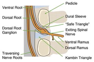
The safe triangle contains epidural fat. The epidural space is contained within the dural sleeve.
Indications and Efficacy
There have been many proposed mechanisms for the effect of epidural steroid injections. They include anti-inflammatory effects, anti-nociceptive effects (blocking of C-fibre transmission[1]), suppression of immune response, mechanical debridement (washing away of inflammatory mediators), stopping the "pain-spasm" cycle, and placebo effect.
The primary indication for transforaminal epidural steroid injection is for lower extremity radicular pain due to lumbar disc herniation in patients:[2]
- who require pain relief
- who have had a failed response to non-surgical intervention or non-surgical interventions aren't indicated
- whose pain likely has an inflammatory basis.
- whose symptoms are consistent with lumbar or sacral radicular pain and may or may not have radiculopathy.
- who have imaging findings that support the diagnosis of radicular pain
Probably the most important efficacy study is that by Ghahreman and colleagues.[3] It was a randomised controlled study where they compared transforaminal steroid with a variety of other control groups. Transforaminal steroid with local anaesthetic resulted in a successful primary outcome 54% of the time and was more effective than the control groups. Success was defined as at least 50% relief one month post injection.
A 2013 systematic review looked at 6 RCTs, 5 pragmatic comparative trials, and 12 observational trials. They found that up to 70% achieve 50% or more greater pain relief, they reduce health care consumption, are surgery sparing, are cost effective, and can be durable with 25-40% having 12 months of relief.[4]
Epidural steroid injection is less effective for spinal stenosis. For example in one 2004 study of mixed caudal/transforaminal patients only around one third having relief of greater than 2 months, one third having relief of less than 2 months, and one third having no relief. There was an overall small magnitude of change.[5] However a pivotal 2014 RCT showed that there was no difference for pain or disability at 6 weeks between the steroid-anaesthetic group and anaesthetic only group.[6]
Contraindications
Contraindications as outlined by the SIS guidelines are:[2]
- Absolute contraindications
- Informed consent not possible
- Not possible to use contrast medium
- Untreated local infection
- Patient non-cooperation despite consent.
- Pregnancy due to fluoroscopic use
- Relative contraindications
- Coagulopathy
- Anatomical abnormalities precluding safe intervention
- Systemic infection
- Significant cardiorespiratory compromise
- Immunosuppression
Pre-procedural Evaluation
Equipment
Most steroid preparations contain preservatives (polyethylene glycol and/or benzyl alcohol), however the clinical effect seems undetectable. All particular steroids have larger particles than RBCs, with betamethasone being the smallest. Dexamethasone is a solution with no particles and is the preferred agent used in New Zealand.
Technique
Fluoroscopy Guided
Subpedicular injection is the standard technique for L1-2 through to L5-S1
- Target visualisation
- AP view through L4-5 disc space. Target position under pedicle identified just below 6 o' clock. However the AP view does not offer an unobstructed needle path
- Ipsilateral oblique (~25 degrees, exact amount depends on patient anatomy and level). Unobstructed needle path is visualised.
- Needle placement
- The needle insertion is marked In the ipsilateral oblique view, under the pedicle. Needle placement is down the beam approaching the target.
- Check AP to confirm needle placement at or just lateral to the 6 o clock pedicle position.
- Check lateral to confirm needle placement just posterior to vertebral body, adjacent to the caudal border of the pedicle above the target nerve.
- Injection
- Inject contrast under live fluoroscopy, using digital subtraction if available.
- Watch for intravascular or intrathecal flow. Abort the procedure if intra-arterial or intrathecal flow seen.
- If flow is suboptimal then adjust the needle position.
- Following contrast inject local anaesthetic followed by steroid. 3mL will cover both the superior and inferior intervertebral discs of the corresponding level of injection 88% of the time.
S1 transforaminal injection is used for S1 radicular pain, most commonly due to L5-S1 paracentral disc herniation. It requires placing the needle through the posterior S1 foramen onto the pedicle of S1.
- Target visualisation
- AP view through L5-S1 disc space, with a cephalo-caudad tilt
- Slight ipsilateral oblique, superimposing the anterior and posterior S1 foramina.
- The target point lies on the caudal border of the S1 pedicle, just dorsal to the internal opening of the S1 anterior sacral foramen.
- Needle placement
- The needle insertion point is marked at the lateral margin of the posterior foramen.
- Advance the needle towards the dorsal surface of the sacrum at the lateral margin of the S1 posterior foramen
- When the needle hits the sacrum, it is readjusted in order to pass superiorly and slightly medially into the S1 posterior sacral foramen. The aim is to hit the middle of the caudal border of the anterior half of the S1 pedicle.
- The needle shouldn't go medial tot he middle of the caudal border of the pedicle.
- Injection
- Inject contrast under live fluoroscopy, using digital subtraction if available.
- Watch for intravascular or intrathecal flow. Intravascular uptake is more common here.
- After contrast inject local anaesthetic followed by steroid. 3mL will cover the L5-S1 disc 92% of the time.
Ultrasound Guided
A cadaver study found that ultrasound guided transforaminal injection was almost as accurate as fluoroscopic guidance,[7] however it is not ready for prime time.
Complications
Aftercare
The injection may be repeated.
Videos
See Also
External Links
References
- ↑ Siddall PJ, Cousins MJ. Spinal pain mechanisms. Spine (Phila Pa 1976). 1997 Jan 1;22(1):98-104. doi: 10.1097/00007632-199701010-00016. PMID: 9122790.
- ↑ 2.0 2.1 Bogduk N. Practice Guidelines for Spinal Diagnostic and Treatment Procedures, 2nd Ed. San Francisco: ISIS; 2013
- ↑ Ghahreman A, Ferch R, Bogduk N. The efficacy of transforaminal injection of steroids for the treatment of lumbar radicular pain. Pain Med. 2010 Aug;11(8):1149-68. doi: 10.1111/j.1526-4637.2010.00908.x. PMID: 20704666.
- ↑ MacVicar J, King W, Landers MH, Bogduk N. The effectiveness of lumbar transforaminal injection of steroids: a comprehensive review with systematic analysis of the published data. Pain Med. 2013 Jan;14(1):14-28. doi: 10.1111/j.1526-4637.2012.01508.x. Epub 2012 Oct 30. PMID: 23110347.
- ↑ Delport EG, Cucuzzella AR, Marley JK, Pruitt CM, Fisher JR. Treatment of lumbar spinal stenosis with epidural steroid injections: a retrospective outcome study. Arch Phys Med Rehabil. 2004 Mar;85(3):479-84. doi: 10.1016/s0003-9993(03)00472-6. PMID: 15031837.
- ↑ Friedly JL, Comstock BA, Turner JA, Heagerty PJ, Deyo RA, Sullivan SD, Bauer Z, Bresnahan BW, Avins AL, Nedeljkovic SS, Nerenz DR, Standaert C, Kessler L, Akuthota V, Annaswamy T, Chen A, Diehn F, Firtch W, Gerges FJ, Gilligan C, Goldberg H, Kennedy DJ, Mandel S, Tyburski M, Sanders W, Sibell D, Smuck M, Wasan A, Won L, Jarvik JG. A randomized trial of epidural glucocorticoid injections for spinal stenosis. N Engl J Med. 2014 Jul 3;371(1):11-21. doi: 10.1056/NEJMoa1313265. Erratum in: N Engl J Med. 2014 Jul 24;371(4):390. PMID: 24988555.
- ↑ Hashemi, Masoud et al. “Ultrasound-Guided Lumbar Transforaminal Epidural Injections; A Single Center Fluoroscopic Validation Study.” Bulletin of emergency and trauma vol. 7,3 (2019): 251-255. doi:10.29252/beat-070307
Literature Review
- Reviews from the last 7 years: review articles, free review articles, systematic reviews, meta-analyses, NCBI Bookshelf
- Articles from all years: PubMed search, Google Scholar search.
- TRIP Database: clinical publications about evidence-based medicine.
- Other Wikis: Radiopaedia, Wikipedia Search, Wikipedia I Feel Lucky, Orthobullets,


