Popliteus Tendinopathy

| |
| Popliteus Tendinopathy | |
|---|---|
| Synonym | Popliteus tendinitis |
The popliteus may be affected by tenosynovitis, acute calcific tendonitis, rupture, and avulsion. Popliteus tendinopathy is rare but often misdiagnosed.
Anatomy
- Main article: Popliteus
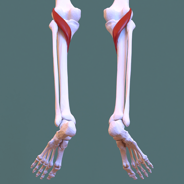
The popliteus muscle has up to three origins - the lateral femoral condyle (just anterior and inferior to the lateral collateral ligament), the fibula head, and in some people also the posterior horn of the lateral meniscus. It inserts into the posterior surface of the proximal tibia proximal to the soleal line.
The muscle is unusual is that its proximal attachment (origin) is tendinous and the muscle belly lies distally. The function of popliteus is internal rotation of the tibia on the femur if the femur is fixed (sitting down) or external rotation of the femur on the tibia if the tibia is fixed (standing up). Along with popliteus, the popliteo-fibular ligament is also important for preventing external tibial rotation. When the knee is in full extension, the femur slightly medially rotates on the tibia to lock the knee joint in place. It unlocks the knee with medial tibial rotation to allow flexion. It also helps to prevent anterior dislocation of the femur / posterior dislocation of the tibia while crouching.
It is supplied by the tibial nerve (L5 and S1) and popliteal artery.
Risk Factors
- Downhill running or other deceleration activities
Clinical Features
Pain is usually felt posterolaterally. Patients may find it difficult to bend their knee. There may be localised tenderness over the popliteus tendon.
There are two special tests.
- Garrick test: patient supine, hip and knee flexed to 90 degrees, leg internally rotated, patient asked to keep the leg there while the examiner passively externally rotates the tibia.
- Passive external rotation: In the same position as above apply passive external force.
Imaging
Calcific changes may be apparent on plan x-ray. MRI may show tendinopathic changes and tenosynovitis.
Treatment
Anecdotally the condition responds well to corticosteroid injection.
Physical therapy is targeted at eccentric strengthening of the quadriceps to reduce strain on the popliteus.
References
Literature Review
- Reviews from the last 7 years: review articles, free review articles, systematic reviews, meta-analyses, NCBI Bookshelf
- Articles from all years: PubMed search, Google Scholar search.
- TRIP Database: clinical publications about evidence-based medicine.
- Other Wikis: Radiopaedia, Wikipedia Search, Wikipedia I Feel Lucky, Orthobullets,


