Shoulder Biomechanics: Difference between revisions
(Created page with "{{stub}} The shoulder girdle links the upper extremity to the trunk. Working with the elbow, it allows the hand to be positioned in space. It is the most dynamic and mobile jo...") |
No edit summary |
||
| Line 1: | Line 1: | ||
{{stub}} | {{stub}} | ||
The shoulder girdle links the upper extremity to the trunk. Working with the elbow, it allows the hand to be positioned in space. It | The shoulder girdle links the upper extremity to the trunk. Working with the elbow, it allows the hand to be positioned in space. It needs a high level of structural protection and functional control. The biomechanics of the shoulder are quite complex. | ||
== Shoulder Complex Structure == | |||
The shoulder complex (shoulder girdle) consists of four joints: glenohumeral (GHJ), acromioclavicular (ACJ), [[Sternoclavicular Joint Anatomy|sternoclavicular]] (SCJ), scapulothoracic joint (STJ). Some individuals also have a coracoclavicular joint (CCJ). These joints need to have free motion and coordinated action, and motion occurs in all of them with any significant arm movement. | |||
=== Sternoclavicular Joint === | |||
[[File:SCJ Gray.png|thumb|The sternoclavicular joint and stabilising structures.]] | |||
{{Main|Sternoclavicular Joint Anatomy}} | |||
The SCJ is formed by the articulations of the proximal clavicle with the clavicular notch of the manubrium and with the cartilage of the first rib. It is a saddle shaped joint with a fibrocartilaginous disc. Stability of the SCJ is provided by very strong ligaments, including the sternoclavicular, costoclavicular, and interclavicular ligaments, as well as by the disc, capsule, and muscular attachments. The SCJ provides the major axis of rotation for motion of the clavicle and scapula. The close-packed position is with maximal shoulder elevation. | |||
=== Acromioclavicular Joint === | |||
[[File:ACJ Gray.png|thumb|Left shoulder: acromioclavicular joint, and anterior ligaments]] | |||
{{Main|Acromioclavicular Joint Anatomy}} | |||
The ACJ if formed by the articulation of the acromion process of the scapula with the distal clavicle. It is an irregular diarthrodial joint. Stability is provided by the coracoclavicular ligaments. There are many variations of the ACJ. It is able to move in all three planes.. Rotation occurs at the ACJ during arm elevation. The close-packed position is with abduction of the humerus to 90 degrees. | |||
=== Coracoclavicular Joint === | |||
This is an anomalous joint formed at the junction of the coracoid process of the scapula and the inferior surface of the clavicle. They are connected by the coracoclavicular ligament. The prevalence is approximately 10%. Some texts describe this structure as normally being a syndesmosis that doesn't permit much movement. | |||
=== Glenohumeral Joint === | |||
{{Main|Glenohumeral Joint Anatomy}} | |||
[[File:Shoulder joint anterior view.png|thumb|Anterior view of the right shoulder rotator cuff muscles]] | |||
[[File:Shoulder joint posterior view.png|thumb|Posterior view of the right shoulder rotator cuff muscles]] | |||
The GHJ is the ball-and-socket articulation between the humeral head and the glenoid fossa of the scapula. The GHJ is the most dynamic and mobile joint in the body. | |||
The head of the GHJ is almost hemispherical and has three to four times the surface area as compared to the shallow glenoid fossa allowing a wide range of rotation. The glenoid fossa also has a less curved shape compared to the humeral head, and this allows for sliding movements. Variations in shape of the glenoid fossa cavity include oval-shaped, egg-shaped, and pear-shaped. | |||
The glenoid labrum is a dense fibrous and fibrocartilagenous tissue that encircles the glenoid fossa. It is formed by the capsule of the joint, the tendon of the long head of biceps brachii, and the glenohumeral ligaments. It is triangular in cross-section. The function is to deepen the fossa and add stability to the joint. | |||
There are several ligaments that merge with the glenohumeral joint capsule: superior, middle, and inferior glenohumeral ligaments on the anterior side; and coracohumeral ligament on the superior side. | |||
The tendons of the four rotator cuff muscles join the joint capsule and form a collagenous cuff around the GHJ surrounding the posterior, superior, and anterior sides. They contribute to rotation of the humerus. The muscles are supraspinatus, infraspinatus, teres minor, and subscapularis. The external rotators are the supraspinatus, infraspinatus, and teres minor. Subscapularis contributes to internal rotation. The fibres of the lateral rotators mingle with one another increasing their tension and functional power. | |||
The tension in the rotator cuff muscles pulls the head of the humerus towards the glenoid fossa, increasing joint stability. Prior to motion of the humerus, tension is developed in the rotator cuff and biceps tendons. There is also negative pressure within the capsule of the GHJ which increases stability. | |||
The close-packed position is with the humerus abducted and externally rotated. | |||
=== Scapulothoracic Joint === | |||
The STJ refers to the region between the anterior scapula and the thoracic wall. The muscles that attach to the scapula include levator scapula, trapezius, and rhomboids. They contract to stabilise the shoulder joint, and facilitate movement of the limb with by appropriately positioning the GHJ. | |||
=== Bursae === | |||
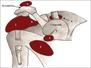
[[File:Bursae shoulder joint.jpg|thumb|Bursae of the shoulder joint: (1) subacromial-subdeltoid bursa, (2) subscapular recess, (3) subcoracoid bursa, (4) coracoclavicular bursa, (5) supra-acromial bursa and (6) medial extension of subacromial-subdeltoid bursa.]] | |||
There are several bursae that reduce friction between layers of tissue and these include the subscapularis, subcoracoid, and subacromial bursae. The most well known, the subacromial bursa, sits in the subacromial space between the acromion and the coracoacromial ligament above and the glenohumeral joint below. It cushions the supraspinatus from the overlying acromion. The subscapularis and subcoracoid bursae are sometimes merged. They reduce friction of the superficial fibres of subscapularis against the neck of the scapula, the head of the humerus, and the coracoid process. | |||
[[Category:Biomechanics]] | [[Category:Biomechanics]] | ||
Revision as of 17:19, 16 August 2021
The shoulder girdle links the upper extremity to the trunk. Working with the elbow, it allows the hand to be positioned in space. It needs a high level of structural protection and functional control. The biomechanics of the shoulder are quite complex.
Shoulder Complex Structure
The shoulder complex (shoulder girdle) consists of four joints: glenohumeral (GHJ), acromioclavicular (ACJ), sternoclavicular (SCJ), scapulothoracic joint (STJ). Some individuals also have a coracoclavicular joint (CCJ). These joints need to have free motion and coordinated action, and motion occurs in all of them with any significant arm movement.
Sternoclavicular Joint
- Main article: Sternoclavicular Joint Anatomy
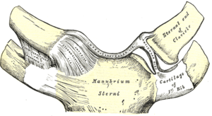
The SCJ is formed by the articulations of the proximal clavicle with the clavicular notch of the manubrium and with the cartilage of the first rib. It is a saddle shaped joint with a fibrocartilaginous disc. Stability of the SCJ is provided by very strong ligaments, including the sternoclavicular, costoclavicular, and interclavicular ligaments, as well as by the disc, capsule, and muscular attachments. The SCJ provides the major axis of rotation for motion of the clavicle and scapula. The close-packed position is with maximal shoulder elevation.
Acromioclavicular Joint
- Main article: Acromioclavicular Joint Anatomy
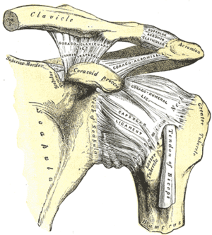
The ACJ if formed by the articulation of the acromion process of the scapula with the distal clavicle. It is an irregular diarthrodial joint. Stability is provided by the coracoclavicular ligaments. There are many variations of the ACJ. It is able to move in all three planes.. Rotation occurs at the ACJ during arm elevation. The close-packed position is with abduction of the humerus to 90 degrees.
Coracoclavicular Joint
This is an anomalous joint formed at the junction of the coracoid process of the scapula and the inferior surface of the clavicle. They are connected by the coracoclavicular ligament. The prevalence is approximately 10%. Some texts describe this structure as normally being a syndesmosis that doesn't permit much movement.
Glenohumeral Joint
- Main article: Glenohumeral Joint Anatomy
The GHJ is the ball-and-socket articulation between the humeral head and the glenoid fossa of the scapula. The GHJ is the most dynamic and mobile joint in the body.
The head of the GHJ is almost hemispherical and has three to four times the surface area as compared to the shallow glenoid fossa allowing a wide range of rotation. The glenoid fossa also has a less curved shape compared to the humeral head, and this allows for sliding movements. Variations in shape of the glenoid fossa cavity include oval-shaped, egg-shaped, and pear-shaped.
The glenoid labrum is a dense fibrous and fibrocartilagenous tissue that encircles the glenoid fossa. It is formed by the capsule of the joint, the tendon of the long head of biceps brachii, and the glenohumeral ligaments. It is triangular in cross-section. The function is to deepen the fossa and add stability to the joint.
There are several ligaments that merge with the glenohumeral joint capsule: superior, middle, and inferior glenohumeral ligaments on the anterior side; and coracohumeral ligament on the superior side.
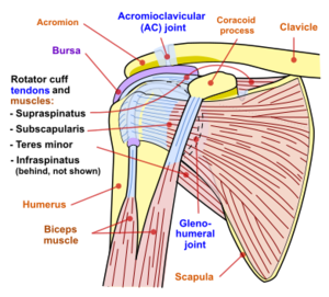
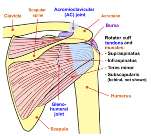
The tendons of the four rotator cuff muscles join the joint capsule and form a collagenous cuff around the GHJ surrounding the posterior, superior, and anterior sides. They contribute to rotation of the humerus. The muscles are supraspinatus, infraspinatus, teres minor, and subscapularis. The external rotators are the supraspinatus, infraspinatus, and teres minor. Subscapularis contributes to internal rotation. The fibres of the lateral rotators mingle with one another increasing their tension and functional power.
The tension in the rotator cuff muscles pulls the head of the humerus towards the glenoid fossa, increasing joint stability. Prior to motion of the humerus, tension is developed in the rotator cuff and biceps tendons. There is also negative pressure within the capsule of the GHJ which increases stability.
The close-packed position is with the humerus abducted and externally rotated.
Scapulothoracic Joint
The STJ refers to the region between the anterior scapula and the thoracic wall. The muscles that attach to the scapula include levator scapula, trapezius, and rhomboids. They contract to stabilise the shoulder joint, and facilitate movement of the limb with by appropriately positioning the GHJ.
Bursae
There are several bursae that reduce friction between layers of tissue and these include the subscapularis, subcoracoid, and subacromial bursae. The most well known, the subacromial bursa, sits in the subacromial space between the acromion and the coracoacromial ligament above and the glenohumeral joint below. It cushions the supraspinatus from the overlying acromion. The subscapularis and subcoracoid bursae are sometimes merged. They reduce friction of the superficial fibres of subscapularis against the neck of the scapula, the head of the humerus, and the coracoid process.
