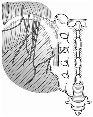
Superior Cluneal Nerve Entrapment
Some authors posit that cluneal nerve entrapment is an under-diagnosed cause of chronic low back and leg pain, and it can cause neuropathic type symptoms. Superior cluneal nerve entrapment is thought to be more common than middle cluneal nerve entrapment.
Anatomy and Pathophysiology

Superior cluneal nerve (SCN) entrapment occurs where the nerve pierces the thoracolumbar attachment at the posterior iliac crest. The medial branch of the SCN consistently passes through an osteofibrous tunnel and can be spontaneously entrapped.
The middle cluneal nerve (MCN) is comprised of the sensory branches of the S1 to S3 dorsal rami. It travels below the PSIS in an approximately horizontal route and supplies the skin overlying the posteromedial buttock. The S1 to S2 composition could explain why some patients may get leg symptoms. It can become trapped with passing underneith or through the long posterior sacroiliac ligament. MCN entrapment can be confused with sacroiliac joint pain. Even a positive sacroiliac joint block could still indicate LPSL pain or MCN entrapment if there is dorsal extravasation of the local anaesthetic.
Epidemiology
In one retrospective review of 834 patient with LBP, 14% were thought to be due to superior cluneal nerve entrapment. See the diagnosis section below for their diagnostic method.[2]
Clinical Features
In those with SCN disorder, around 50% have leg symptoms, but only 1% have leg symptoms only. The most commonly aggravating features are walking, rising from sitting, and standing. The prevalence of vertebral compression fractures is higher in those with SCN than in those without SCN disorder (26% vs 12%), most commonly at T12-L3.[2]
Diagnosis
- Superior cluneal nerve entrapment
There is no agreed diagnostic method. The diagnosis can be considered by tenderness over the iliac crest or LPSL causing provocation of symptoms. Pain relief following local anaesthetic injection can give further weight to the diagnosis.
Hiroshi et al used the following diagnostic criteria. [2]
- Maximal tender point on posterior iliac crest approximately 70 mm from the midline and 45 mm from the PSIS where the medial branch of the SCN runs through an osteofibrous tunnel consisting of the thoraco-lumbar fascia and the iliac crest and
- Palpation of the maximally tender point reproduced the chief complaint of LBP and/or leg symptoms
In their study, when patients met both criteria, a nerve block injection was performed. If relief of symptoms is obtained following injection that can give further weight to the diagnosis. In their study, if repeated injections failed to relieve symptoms then they had SCN decompression.
- Middle cluneal nerve entrapment
Again there is no agreed diagnostic method. The MCN tender point is typically on the LPSL within 40mm caudal to the PSIS.
Treatment
Injections
Trigger point injections have been described.
Surgery
Reports have been published on SCN release and MCN release. Most reports are on SCN release.
References
- ↑ Aota. Entrapment of middle cluneal nerves as an unknown cause of low back pain. World journal of orthopedics 2016. 7:167-70. PMID: 27004164. DOI. Full Text.
- ↑ 2.0 2.1 2.2 Kuniya et al.. Prospective study of superior cluneal nerve disorder as a potential cause of low back pain and leg symptoms. Journal of orthopaedic surgery and research 2014. 9:139. PMID: 25551470. DOI. Full Text.
Literature Review
- Reviews from the last 7 years: review articles, free review articles, systematic reviews, meta-analyses, NCBI Bookshelf
- Articles from all years: PubMed search, Google Scholar search.
- TRIP Database: clinical publications about evidence-based medicine.
- Other Wikis: Radiopaedia, Wikipedia Search, Wikipedia I Feel Lucky, Orthobullets,


