Transitional Vertebral Anatomy
See Konin et al for an open access review on the lumbosacral transitional vertebral (LSTV) anatomy.[1]
Numbering Technique
It can be difficult to establish whether a LSTV is a lumbarised S1 or a sacralised L5, and there have been various techniques described, with the best technique being high quality imaging of the entire spine.[1]
Survey sequences
In New Zealand, some private radiology providers will do a sagittal survey sequence of the entire spine (also called a localiser) as routine while others do not. You then count from the odontoid peg of C2 inferiorly, rather than counting cranially from L5. This also allows one to differentiate between the hypoplastic rib and the lumbar transverse process. Therefore one can next correctly identify the L1 vertebral body, and subsequently determine the correct numeric assignment of the LSTV. Radiographs of the entire spine can fulfil the same purpose. If there is only a normal lumbar spine radiograph, then correct numeration may still be possible, but it can be tricky at the thoracolumbar junction to differentiate the hypoplastic rib from the transverse process. Thoracolumbar transitions can also make things difficult without a full spine view.[1]
Using a sagittal localiser sequence assumes there are 7 cervical and 12 thoracic vertebra. The method does not account for thoracolumbar transitional anatomy or allow one to differentiate between dysplastic ribs and lumbar transverse processes. Adding a coronal localising sequence can sometimes increase the accuracy of locating the thoracolumbar junction.[1]
Iliolumbar ligament
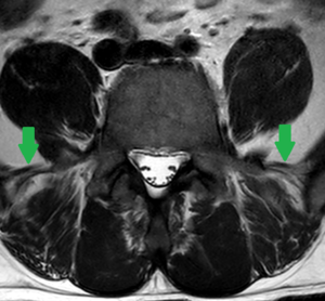
Locating the iliolumbar ligament is another technique described, as this ligament is present at the L5 transverse process in 96.8% of people.[2] On MRI it is a low signal intensity structure on T1 and T2 images and looks like a single or double band that extends from the transverse process of L5 to the posteromedial iliac crest. It serves to restrict flexion, extension, axial rotation, and lateral bending of L5 on S1. In cases of thoracolumbar transitional vertebrae, identification of the iliolumbar ligament is not sufficient to identify the L5 vertebral body.[1]
Other anatomic markers
Using other markers such as the aortic bifurcation, right renal artery, and conus medullaris has also been described and are not satisfactory.[1]
References
- ↑ 1.0 1.1 1.2 1.3 1.4 1.5 Konin & Walz. Lumbosacral transitional vertebrae: classification, imaging findings, and clinical relevance. AJNR. American journal of neuroradiology 2010. 31:1778-86. PMID: 20203111. DOI.
- ↑ Carrino et al.. Effect of spinal segment variants on numbering vertebral levels at lumbar MR imaging. Radiology 2011. 259:196-202. PMID: 21436097. DOI.


