File:Finger extensor mechanism dorsal view.jpg
Finger_extensor_mechanism_dorsal_view.jpg (324 × 532 pixels, file size: 23 KB, MIME type: image/jpeg)
Summary
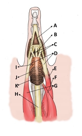
a: triangular ligament; b: central slip; c: slips of long extensor tendon to lateral bands; d: lateral bands; e: lumbrical muscle; f: superficial belly of dorsal interosseous muscle; g: deep belly of dorsal interosseous muscle or palmar interosseous muscle; h: long extensor tendon; i: oblique fibers; j: transverse fibers; k: saggital bands; l: oblique retinacular ligament (Landmeer’s ligament)
Ditsios K, Konstantinou P, Pinto I, Karavelis A, Kostretzis L (2017) Extensor Mechanism’s Anatomy at the Metacarpophalangeal Joint. MOJ Orthop Rheumatol 8(4): 00319. DOI: 10.15406/mojor.2017.08.00319
Licencing
![]()
This work is licensed under the Creative Commons Attribution-NonCommercial 4.0 License.
File history
Click on a date/time to view the file as it appeared at that time.
| Date/Time | Thumbnail | Dimensions | User | Comment | |
|---|---|---|---|---|---|
| current | 12:50, 6 February 2022 | | 324 × 532 (23 KB) | Jeremy (talk | contribs) | a: triangular ligament; b: central slip; c: slips of long extensor tendon to lateral bands; d: lateral bands; e: lumbrical muscle; f: superficial belly of dorsal interosseous muscle; g: deep belly of dorsal interosseous muscle or palmar interosseous muscle; h: long extensor tendon; i: oblique fibers; j: transverse fibers; k: saggital bands; l: oblique retinacular ligament (Landmeer’s ligament) Ditsios K, Konstantinou P, Pinto I, Karavelis A, Kostretzis L (2017) Extensor Mechanism’s Anatomy a... |
You cannot overwrite this file.
File usage
The following 3 pages use this file:

