File:Myotendinous junction structure.png

Original file (850 × 648 pixels, file size: 137 KB, MIME type: image/png)
Summary
Structure of the myotendinous junction. (A) Transmission electron microscopy image of myofibrils (scale bar = 2 µm) (B) Transmission electron microscopy of the MTJ of the rat sternomastoid muscle (M = muscle side; T = Tendon side) (scale bar = 0.5 µm) (C) View of the tendon fibres observed with SEM (scale bar = 1.8 µm) (D) Graphical representation of the MTJ and its components
Modified (corrected errors) from Sensini, Alberto et al. “Tissue Engineering for the Insertions of Tendons and Ligaments: An Overview of Electrospun Biomaterials and Structures.” Frontiers in bioengineering and biotechnology vol. 9 645544. 2 Mar. 2021, doi:10.3389/fbioe.2021.645544
Licencing
![]()
This work is licensed under a Creative Commons Attribution 4.0 International License.
File history
Yi efo/eka'e gwa ebo wo le nyangagi wuncin ye kamina wunga tinya nan
| Gwalagizhi | Nyangagi | Dimensions | User | Comment | |
|---|---|---|---|---|---|
| current | 20:16, 9 August 2021 | | 850 × 648 (137 KB) | Jeremy (talk | contribs) | Corrected some errors |
| 18:41, 9 August 2021 | 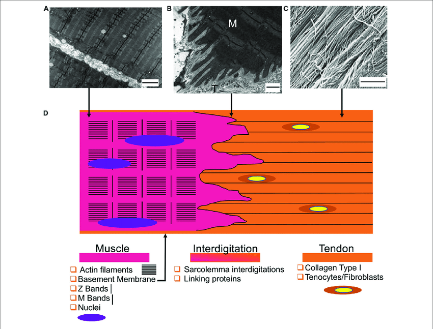 | 850 × 647 (139 KB) | Jeremy (talk | contribs) | From Sensini, Alberto et al. “Tissue Engineering for the Insertions of Tendons and Ligaments: An Overview of Electrospun Biomaterials and Structures.” Frontiers in bioengineering and biotechnology vol. 9 645544. 2 Mar. 2021, doi:10.3389/fbioe.2021.645544 |
You cannot overwrite this file.
File usage
The following page uses this file:

