Enthesis: Difference between revisions
No edit summary |
No edit summary |
||
| Line 3: | Line 3: | ||
==Structure and Function== | ==Structure and Function== | ||
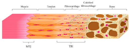
[[File:Enthesis structure.png|thumb|alt=| | [[File:Myotendinous junction and enthesis combined.png|thumb|450px|The [[Myotendinous Junction|myotendinous junction]] (MTJ) and fibrocartilaginous enthesis (tendon‐to‐bone interface: TBI) with its different zones: tendon, fibrocartilage, calcified fibrocartilage and bone<ref>Bianchi E, Ruggeri M, Rossi S, Vigani B, Miele D, Bonferoni MC, Sandri G, Ferrari F. Innovative Strategies in Tendon Tissue Engineering. Pharmaceutics. 2021 Jan 11;13(1):89. doi: 10.3390/pharmaceutics13010089. PMID: 33440840; PMCID: PMC7827834.</ref>|alt=]] | ||
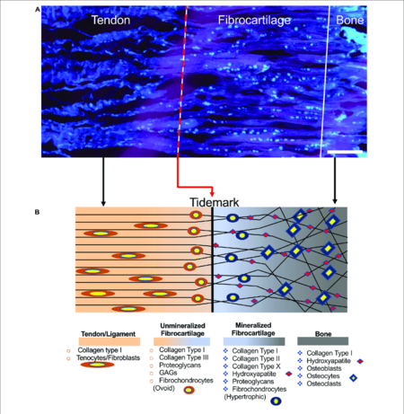
[[File:Enthesis structure.png|thumb|alt=|450px|Structure of the enthesis. (A) Enthesis cryocut section of a porcine Achilles tendon was stained for cells using SYTO R 13. Cells are depicted cyan (scale bar = 150 µm). (B) Graphical representation of the enthesis and its components.<ref>Sensini, Alberto et al. “Tissue Engineering for the Insertions of Tendons and Ligaments: An Overview of Electrospun Biomaterials and Structures.” ''Frontiers in bioengineering and biotechnology'' vol. 9 645544. 2 Mar. 2021, doi:10.3389/fbioe.2021.645544</ref>]] | |||
The musculoskeletal system as a whole has both hard and soft tissues. The interfaces between these tissues have gradients in order to reduce stress concentrations at the junction sites. The interfaces between tendons/ligaments and bone are called entheses, while the interfaces between tendons and muscles are called myotendinous junctions (MTJ). | |||
The structure of the enthesis varies widely depending on the location. There are two main categories of entheses: the indirect fibrous enthesis, and the direct fibrocartilaginous enthesis. | The structure of the enthesis varies widely depending on the location. There are two main categories of entheses: the indirect fibrous enthesis, and the direct fibrocartilaginous enthesis. | ||
Revision as of 19:58, 9 August 2021
The enthesis (plural entheses) is the interface between tendon/ligament and bone. See also about the myotendinous junction, the connection between muscle and tendon.
Structure and Function


The musculoskeletal system as a whole has both hard and soft tissues. The interfaces between these tissues have gradients in order to reduce stress concentrations at the junction sites. The interfaces between tendons/ligaments and bone are called entheses, while the interfaces between tendons and muscles are called myotendinous junctions (MTJ).
The structure of the enthesis varies widely depending on the location. There are two main categories of entheses: the indirect fibrous enthesis, and the direct fibrocartilaginous enthesis.
The function of the enthesis is to dissipate stress away from the interface.
Fibrous Enthesis
The fibrous enthesis consists of tendons and ligaments being connected through acute angles to bones with collagen fibres extending directly from the periosteum, termed Sharpey's fibres.
Fibrocartilaginous Enthesis
The fibrocartilaginous enthesis consists of a progressive mineralisation gradient that is organised into four zones. The boundary between the unmineralised and mineralised fibrocartilage zones is called the tidemark where there is an abrupt transition. The thickness of the junction is around 500 µm.
- Tendon/ligament: longitudinally oriented fibroblasts and a parallel arrangement of collagen fibres.
- Unmineralised fibrocartilage: contains various collagens (types I, II, III, X, IX) and proteoglycans (mostly aggrecans with associated chondroitin 4- and 6- glycosaminoglycans). The collagen fibres become increasingly randomly arranged. Fibroblasts and tenocytes are replaced by ovoid-shaped aligned fibrochondrocytes.
- Mineralised fibrocartilage. There are hypertrophic chondrocytes surrounded by type II and X collagens and aggrecans.
- Bone
Clinical Applications
A disease process affecting the entheses is called enthesopathy or enthesitis.
Common locations for injury are the rotator cuff, the anterior cruciate ligament, the Achilles tendon, the medial collateral ligament of the knee, tennis elbow, and jumper's knee.
Enthesopathy is also associated with spondyloarthropathies.
References
- ↑ Bianchi E, Ruggeri M, Rossi S, Vigani B, Miele D, Bonferoni MC, Sandri G, Ferrari F. Innovative Strategies in Tendon Tissue Engineering. Pharmaceutics. 2021 Jan 11;13(1):89. doi: 10.3390/pharmaceutics13010089. PMID: 33440840; PMCID: PMC7827834.
- ↑ Sensini, Alberto et al. “Tissue Engineering for the Insertions of Tendons and Ligaments: An Overview of Electrospun Biomaterials and Structures.” Frontiers in bioengineering and biotechnology vol. 9 645544. 2 Mar. 2021, doi:10.3389/fbioe.2021.645544

