Enthesis
The enthesis is the interface between tendon/ligament and bone. See also about the myotendinous junction, the connection between muscle and tendon.
Structure and Function
The structure of the enthesis varies widely depending on the location. There are two main categories of entheses: the indirect fibrous enthesis, and the direct fibrocartilaginous enthesis.
The fibrous enthesis consists of tendons and ligaments being connected through acute angles to bones with collagen fibres extending directly from the periosteum, termed Sharpey's fibres.
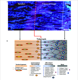
The fibrocartilaginous enthesis consists of a progressive mineralisation gradient that is organised into four zones. The boundary between the unmineralised and mineralised fibrocartilage zones is called the tidemark. The thickness of the junction is around 500 µm.
- Tendon/ligament
- Unmineralised fibrocartilage: contains various collagens (types I, II, III, X, IX) and proteoglycans (mostly aggrecans with associated chondroitin 4- and 6- glycosaminoglycans). The collagen fibres become increasingly randomly arranged. Fibroblasts and tenocytes are replaced by ovoid-shaped aligned fibrochondrocytes.
- Mineralised fibrocartilage. There are hypertrophic chondrocytes surrounded by type II and X collagens and aggrecans.
- Bone
The function of the enthesis is to dissipate stress away from the interface.
Clinical Applications
- Injury
Common locations for injury are the rotator cuff, the anterior cruciate ligament, the Achilles tendon, the medial collateral ligament of the knee, tennis elbow, and jumper's knee.


